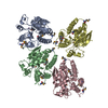
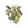
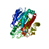
Deposited unit
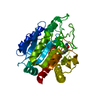
A: Metallophos domain-containing protein
B: Metallophos domain-containing protein
C: Metallophos domain-containing protein
D: Metallophos domain-containing protein
hetero molecules Summary Component details
Theoretical mass Number of molelcules Total (without water) 145,959 12 Polymers 145,516 4 Non-polymers 443 8 Water 4,486 249
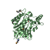
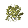
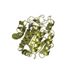
1
A: Metallophos domain-containing protein
hetero molecules Summary Component details Symmetry operations
Theoretical mass Number of molelcules Total (without water) 36,490 3 Polymers 36,379 1 Non-polymers 111 2 Water 18 1
Type Name Symmetry operation Number identity operation 1_555 x,y,z 1
2
B: Metallophos domain-containing protein
hetero molecules Summary Component details Symmetry operations
Theoretical mass Number of molelcules Total (without water) 36,490 3 Polymers 36,379 1 Non-polymers 111 2 Water 18 1
Type Name Symmetry operation Number identity operation 1_555 x,y,z 1
3
C: Metallophos domain-containing protein
hetero molecules Summary Component details Symmetry operations
Theoretical mass Number of molelcules Total (without water) 36,490 3 Polymers 36,379 1 Non-polymers 111 2 Water 18 1
Type Name Symmetry operation Number identity operation 1_555 x,y,z 1
4
D: Metallophos domain-containing protein
hetero molecules Summary Component details Symmetry operations
Theoretical mass Number of molelcules Total (without water) 36,490 3 Polymers 36,379 1 Non-polymers 111 2 Water 18 1
Type Name Symmetry operation Number identity operation 1_555 x,y,z 1
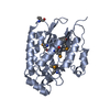
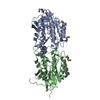
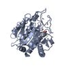
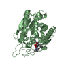
5
A: Metallophos domain-containing protein
hetero molecules
B: Metallophos domain-containing protein
hetero molecules Summary Component details Symmetry operations Calculated values
Theoretical mass Number of molelcules Total (without water) 72,980 6 Polymers 72,758 2 Non-polymers 222 4 Water 36 2
Type Name Symmetry operation Number identity operation 1_555 x,y,z 1 crystal symmetry operation 3_655 -x+1,y+1/2,-z+1/2 1
Buried area 2760 Å2 ΔGint -52 kcal/mol Surface area 22450 Å2 Method
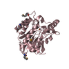
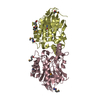
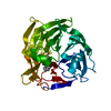
6
C: Metallophos domain-containing protein
hetero molecules
D: Metallophos domain-containing protein
hetero molecules Summary Component details Symmetry operations Calculated values
Theoretical mass Number of molelcules Total (without water) 72,980 6 Polymers 72,758 2 Non-polymers 222 4 Water 36 2
Type Name Symmetry operation Number identity operation 1_555 x,y,z 1 crystal symmetry operation 4_545 x+1/2,-y-1/2,-z 1
Buried area 2910 Å2 ΔGint -52 kcal/mol Surface area 22240 Å2 Method
Unit cell Length a, b, c (Å) 74.190, 92.534, 183.626 Angle α, β, γ (deg.) 90.000, 90.000, 90.000 Int Tables number 19 Space group name H-M P21 21 21 Space group name Hall P2ac2ab Symmetry operation #1 : x,y,z#2 : x+1/2,-y+1/2,-z#3 : -x,y+1/2,-z+1/2#4 : -x+1/2,-y,z+1/2
Noncrystallographic symmetry (NCS) NCS domain NCS domain segments Ens-ID
Show large table (11 x 24) Hide large table Dom-ID Component-ID Beg auth comp-ID Beg label comp-ID End auth comp-ID End label comp-ID Selection details Auth asym-ID Label asym-ID Auth seq-ID Label seq-ID 1 1 GLYGLYPROPRO(chain 'A' and (resid 15 through 30 or (resid 31...AA15 - 92 15 - 92 1 2 GLNGLNASNASN(chain 'A' and (resid 15 through 30 or (resid 31...AA94 - 106 94 - 106 1 3 CYSCYSASPASP(chain 'A' and (resid 15 through 30 or (resid 31...AA108 - 109 108 - 109 1 4 GLYGLYPROPRO(chain 'A' and (resid 15 through 30 or (resid 31...AA112 - 134 112 - 134 1 5 PHEPHEASPASP(chain 'A' and (resid 15 through 30 or (resid 31...AA136 - 198 136 - 198 1 6 GLNGLNPROPRO(chain 'A' and (resid 15 through 30 or (resid 31...AA201 - 290 201 - 290 2 1 GLYGLY
 Movie
Movie Controller

 Yorodumi
 Open data
 Basic information
 Components
 Keywords
 Function and homology information
 Mycobacterium tuberculosis (bacteria)
 X-RAY DIFFRACTION /
  Authors
 United States, 2items
  Citation
 Structure visualization
 Downloads & links
 Other downloads
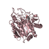

 PDBj
PDBj
 Assembly