 Movie
Movie Controller

[English] 日本語
 Yorodumi
- PDB-6ujf: Crystal structure of the Clostridial cellulose synthase subunit Z... -
+ Open data

- Basic information
| Entry | Database: PDB / ID: 6ujf | ||||||
|---|---|---|---|---|---|---|---|
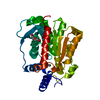
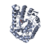
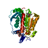
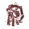
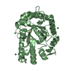
| Title | Crystal structure of the Clostridial cellulose synthase subunit Z (CcsZ) from Clostridioides difficile | ||||||
 Components Components | Endoglucanase | ||||||
 Keywords Keywords | HYDROLASE / glycosyl / biofilm / cellulose / GH8 | ||||||
| Function / homology | : / cellulase / Glycoside hydrolase, family 5 / Cellulase (glycosyl hydrolase family 5) / cellulase activity / membrane => GO:0016020 / Glycoside hydrolase superfamily / beta-D-glucopyranose / Endoglucanase H Function and homology information Function and homology information | ||||||
| Biological species |  Clostridioides difficile (bacteria) Clostridioides difficile (bacteria) | ||||||
| Method |  X-RAY DIFFRACTION / SYNCHROTRON / MOLECULAR REPLACEMENT / Resolution: 2 Å X-RAY DIFFRACTION / SYNCHROTRON / MOLECULAR REPLACEMENT / Resolution: 2 Å | ||||||
 Authors Authors | Scott, W. / Lowrance, B. / Anderson, A.C. / Weadge, J.T. | ||||||
| Funding support |  Canada, 1items Canada, 1items
| ||||||
 Citation Citation | Journal: Plos One / Year: 2020 Title: Identification of the Clostridial cellulose synthase and characterization of the cognate glycosyl hydrolase, CcsZ. Authors: Scott, W. / Lowrance, B. / Anderson, A.C. / Weadge, J.T. | ||||||
| History |
|
- Structure visualization
| Structure viewer | Molecule: MolmilJmol/JSmol |
|---|
- Downloads & links
-Download
| PDBx/mmCIF format | 6ujf.cif.gz | 82.3 KB | Display | PDBx/mmCIF format |
|---|---|---|---|---|
| PDB format | pdb6ujf.ent.gz | 59 KB | Display | PDB format |
| PDBx/mmJSON format | 6ujf.json.gz | Tree view | PDBx/mmJSON format | |
| Others |  Other downloads Other downloads |
-Validation report
| Summary document | 6ujf_validation.pdf.gz | 269 KB | Display | wwPDB validaton report |
|---|---|---|---|---|
| Full document | 6ujf_full_validation.pdf.gz | 269 KB | Display | |
| Data in XML | 6ujf_validation.xml.gz | 1.4 KB | Display | |
| Data in CIF | 6ujf_validation.cif.gz | 4.9 KB | Display | |
| Arichive directory | https://data.pdbj.org/pub/pdb/validation_reports/uj/6ujfftp://data.pdbj.org/pub/pdb/validation_reports/uj/6ujf | HTTPS FTP |
-Related structure data
| Related structure data |  6ujeC  3ujeS S: Starting model for refinement C: citing same article ( |
|---|---|
| Similar structure data |


-Links
 PDBj
PDBj


- Assembly
| Deposited unit | 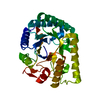
| ||||||||
|---|---|---|---|---|---|---|---|---|---|
| 1 |
| ||||||||
| Unit cell |
|
-Components
| #1: Protein | Mass: 38371.230 Da / Num. of mol.: 1 Source method: isolated from a genetically manipulated source Source: (gene. exp.) Clostridioides difficile (bacteria) / Gene: celH, SAMEA1402348_00819 / Production host: |
|---|---|
| #2: Sugar | ChemComp-BGC /   Type: D-saccharide, beta linking / Mass: 180.156 Da / Num. of mol.: 1 Type: D-saccharide, beta linking / Mass: 180.156 Da / Num. of mol.: 1Source method: isolated from a genetically manipulated source Formula: C6H12O6 |
| #3: Water | ChemComp-HOH /  Mass: 18.015 Da / Num. of mol.: 82 / Source method: isolated from a natural source / Formula: H2O Mass: 18.015 Da / Num. of mol.: 82 / Source method: isolated from a natural source / Formula: H2O |
| Has ligand of interest | N |
| Sequence details | There is no UniProt sequence available at the time of processing. The deposited sequence ...There is no UniProt sequence available at the time of processing. The deposited sequence corresponds to the GenBank entry WP_077724661.1. |
-Experimental details
-Experiment
| Experiment | Method: X-RAY DIFFRACTION / Number of used crystals: 1 |
|---|
- Sample preparation
| Crystal | Density Matthews: 2.11 Å3/Da / Density % sol: 41.58 % |
|---|---|
| Crystal grow | Temperature: 290 K / Method: vapor diffusion, hanging drop / pH: 6 Details: 100 mM MES, 200 mM calcium acetate, 20% (v/v) poly(ethylene glycol) 8000, 2.5 mM cellotriose |
-Data collection
| Diffraction | Mean temperature: 100 K / Serial crystal experiment: N |
|---|---|
| Diffraction source | Source: SYNCHROTRON / Site: CLSI / Beamline: 08ID-1 / Wavelength: 0.9795 Å |
| Detector | Type: DECTRIS PILATUS3 S 6M / Detector: PIXEL / Date: Jun 27, 2019 |
| Radiation | Monochromator: ACCEL/BRUKER DCM / Protocol: SINGLE WAVELENGTH / Monochromatic (M) / Laue (L): M / Scattering type: x-ray |
| Radiation wavelength | Wavelength: 0.9795 Å / Relative weight: 1 |
| Reflection | Resolution: 1.9→31.692 Å / Num. obs: 25294 / % possible obs: 99.5 % / Redundancy: 2 % / Biso Wilson estimate: 48.53 Å2 / CC1/2: 1 / Rmerge(I) obs: 0.01846 / Rpim(I) all: 0.01846 / Rrim(I) all: 0.02611 / Net I/σ(I): 17.96 |
| Reflection shell | Resolution: 1.9→2.071 Å / Redundancy: 2 % / Rmerge(I) obs: 0.2836 / Mean I/σ(I) obs: 2.4 / Num. unique obs: 2157 / CC1/2: 0.903 / Rpim(I) all: 0.2836 / Rrim(I) all: 0.4011 / % possible all: 99.58 |
- Processing
| Software |
| ||||||||||||||||||||||||||||||||||||||||||||||||||||||
|---|---|---|---|---|---|---|---|---|---|---|---|---|---|---|---|---|---|---|---|---|---|---|---|---|---|---|---|---|---|---|---|---|---|---|---|---|---|---|---|---|---|---|---|---|---|---|---|---|---|---|---|---|---|---|---|
| Refinement | Method to determine structure: MOLECULAR REPLACEMENT Starting model: 3UJE Resolution: 2→31.692 Å / SU ML: 0.32 / Cross valid method: THROUGHOUT / σ(F): 1.35 / Phase error: 37.24 / Stereochemistry target values: ML
| ||||||||||||||||||||||||||||||||||||||||||||||||||||||
| Solvent computation | Shrinkage radii: 0.9 Å / VDW probe radii: 1.11 Å / Solvent model: FLAT BULK SOLVENT MODEL | ||||||||||||||||||||||||||||||||||||||||||||||||||||||
| Displacement parameters | Biso max: 86.87 Å2 / Biso mean: 44.2928 Å2 / Biso min: 21.23 Å2 | ||||||||||||||||||||||||||||||||||||||||||||||||||||||
| Refinement step | Cycle: final / Resolution: 2→31.692 Å
| ||||||||||||||||||||||||||||||||||||||||||||||||||||||
| LS refinement shell | Refine-ID: X-RAY DIFFRACTION / Rfactor Rfree error: 0
|
