 Movie
Movie Controller

[English] 日本語
 Yorodumi
- PDB-6u83: OmpA-like domain of FopA1 from Francisella tularensis subsp. tula... -
+ Open data

- Basic information
| Entry | Database: PDB / ID: 6u83 | ||||||
|---|---|---|---|---|---|---|---|
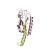
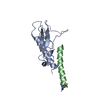
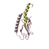
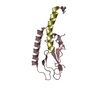
| Title | OmpA-like domain of FopA1 from Francisella tularensis subsp. tularensis SCHU S4 | ||||||
 Components Components | Outer membrane associated protein | ||||||
 Keywords Keywords | PEPTIDE BINDING PROTEIN / outer membrane associated protein / Structural Genomics / Center for Structural Genomics of Infectious Diseases / CSGID | ||||||
| Function / homology |  Function and homology information Function and homology informationporin activity / pore complex / monoatomic ion transport / cell outer membrane Similarity search - Function | ||||||
| Biological species |  Francisella tularensis subsp. tularensis (bacteria) Francisella tularensis subsp. tularensis (bacteria) | ||||||
| Method |  X-RAY DIFFRACTION / SYNCHROTRON / MOLECULAR REPLACEMENT / molecular replacement / Resolution: 2.3566 Å X-RAY DIFFRACTION / SYNCHROTRON / MOLECULAR REPLACEMENT / molecular replacement / Resolution: 2.3566 Å | ||||||
 Authors Authors | Michalska, K. / Skarina, T. / Stogios, P.J. / Di Leo, R. / Savchenko, A. / Joachimiak, A. / Center for Structural Genomics of Infectious Diseases (CSGID) | ||||||
| Funding support |  United States, 1items United States, 1items
| ||||||
 Citation Citation | Journal: To Be Published Title: OmpA-like domain of FopA1 from Francisella tularensis subsp. tularensis SCHU S4 Authors: Michalska, K. / Skarina, T. / Stogios, P.J. / Di Leo, R. / Savchenko, A. / Joachimiak, A. | ||||||
| History |
|
- Structure visualization
| Structure viewer | Molecule: MolmilJmol/JSmol |
|---|
- Downloads & links
-Download
| PDBx/mmCIF format | 6u83.cif.gz | 77.6 KB | Display | PDBx/mmCIF format |
|---|---|---|---|---|
| PDB format | pdb6u83.ent.gz | 56.7 KB | Display | PDB format |
| PDBx/mmJSON format | 6u83.json.gz | Tree view | PDBx/mmJSON format | |
| Others |  Other downloads Other downloads |
-Validation report
| Arichive directory | https://data.pdbj.org/pub/pdb/validation_reports/u8/6u83ftp://data.pdbj.org/pub/pdb/validation_reports/u8/6u83 | HTTPS FTP |
|---|
-Related structure data
| Related structure data |  5u1hS S: Starting model for refinement |
|---|---|
| Similar structure data | |
| Other databases |


-Links
 PDBj
PDBj

- Assembly
| Deposited unit | 
| ||||||||
|---|---|---|---|---|---|---|---|---|---|
| 1 |
| ||||||||
| Unit cell |
|
-Components
| #1: Protein | Mass: 18740.809 Da / Num. of mol.: 1 / Fragment: OmpA-like domain (UNP residues 223-393) Source method: isolated from a genetically manipulated source Source: (gene. exp.) Francisella tularensis subsp. tularensis (strain SCHU S4 / Schu 4) (bacteria)Strain: SCHU S4 / Schu 4 / Gene: fopA1, FTT_0583 / Plasmid: pMCSG53 / Production host: |
|---|---|
| #2: Chemical | ChemComp-EPE /   Mass: 238.305 Da / Num. of mol.: 1 / Source method: obtained synthetically / Formula: C8H18N2O4S / Comment: pH buffer*YM Mass: 238.305 Da / Num. of mol.: 1 / Source method: obtained synthetically / Formula: C8H18N2O4S / Comment: pH buffer*YM |
| #3: Chemical | ChemComp-1PE /   Mass: 238.278 Da / Num. of mol.: 1 / Source method: obtained synthetically / Formula: C10H22O6 / Comment: precipitant*YM Mass: 238.278 Da / Num. of mol.: 1 / Source method: obtained synthetically / Formula: C10H22O6 / Comment: precipitant*YM |
| #4: Chemical | ChemComp-DAL /   Type: D-peptide linking / Mass: 89.093 Da / Num. of mol.: 1 / Source method: obtained synthetically / Formula: C3H7NO2 / Feature type: SUBJECT OF INVESTIGATION Type: D-peptide linking / Mass: 89.093 Da / Num. of mol.: 1 / Source method: obtained synthetically / Formula: C3H7NO2 / Feature type: SUBJECT OF INVESTIGATION |
| #5: Water | ChemComp-HOH /  Mass: 18.015 Da / Num. of mol.: 5 / Source method: isolated from a natural source / Formula: H2O Mass: 18.015 Da / Num. of mol.: 5 / Source method: isolated from a natural source / Formula: H2O |
| Has ligand of interest | Y |
| Has protein modification | Y |
-Experimental details
-Experiment
| Experiment | Method: X-RAY DIFFRACTION / Number of used crystals: 1 |
|---|
- Sample preparation
| Crystal | Density Matthews: 3.2 Å3/Da / Density % sol: 65.51 % |
|---|---|
| Crystal grow | Temperature: 295 K / Method: sitting drop / pH: 7.5 Details: 25% PEG3350, 0.2 M sodium chloride, 0.1 M HEPES, pH 7.5, cryoprotectant: paratone, SITTING DROP, TEMPERATURE 295K |
-Data collection
| Diffraction | Mean temperature: 100 K / Serial crystal experiment: N |
|---|---|
| Diffraction source | Source: SYNCHROTRON / Site: APS / Beamline: 19-ID / Wavelength: 0.9792 Å |
| Detector | Type: DECTRIS PILATUS3 X 6M / Detector: PIXEL / Date: Aug 19, 2019 / Details: mirrors |
| Radiation | Monochromator: double crystal Si(111) / Protocol: SINGLE WAVELENGTH / Monochromatic (M) / Laue (L): M / Scattering type: x-ray |
| Radiation wavelength | Wavelength: 0.9792 Å / Relative weight: 1 |
| Reflection | Resolution: 2.35→50 Å / Num. obs: 10225 / % possible obs: 98.9 % / Observed criterion σ(I): -3 / Redundancy: 11.9 % / Biso Wilson estimate: 55.94 Å2 / CC1/2: 0.999 / Rmerge(I) obs: 0.145 / Net I/σ(I): 26.25 |
| Reflection shell | Resolution: 2.35→2.39 Å / Redundancy: 10.6 % / Rmerge(I) obs: 1.157 / Mean I/σ(I) obs: 1.96 / Num. unique obs: 528 / CC1/2: 0.899 / % possible all: 99.6 |
-Phasing
| Phasing | Method: molecular replacement |
|---|
- Processing
| Software |
| ||||||||||||||||||||||||||||||||||||||||||||||||||||||||||||||||||||||||||||||||||||||||||||||||||||
|---|---|---|---|---|---|---|---|---|---|---|---|---|---|---|---|---|---|---|---|---|---|---|---|---|---|---|---|---|---|---|---|---|---|---|---|---|---|---|---|---|---|---|---|---|---|---|---|---|---|---|---|---|---|---|---|---|---|---|---|---|---|---|---|---|---|---|---|---|---|---|---|---|---|---|---|---|---|---|---|---|---|---|---|---|---|---|---|---|---|---|---|---|---|---|---|---|---|---|---|---|---|
| Refinement | Method to determine structure: MOLECULAR REPLACEMENT Starting model: PDB entry 5U1H Resolution: 2.3566→43.5059 Å / SU ML: 0.26 / Cross valid method: THROUGHOUT / σ(F): 1.35 / Phase error: 36.98 / Stereochemistry target values: ML
| ||||||||||||||||||||||||||||||||||||||||||||||||||||||||||||||||||||||||||||||||||||||||||||||||||||
| Solvent computation | Shrinkage radii: 0.9 Å / VDW probe radii: 1.11 Å / Solvent model: FLAT BULK SOLVENT MODEL | ||||||||||||||||||||||||||||||||||||||||||||||||||||||||||||||||||||||||||||||||||||||||||||||||||||
| Displacement parameters | Biso max: 172.21 Å2 / Biso mean: 82.0066 Å2 / Biso min: 43.62 Å2 | ||||||||||||||||||||||||||||||||||||||||||||||||||||||||||||||||||||||||||||||||||||||||||||||||||||
| Refinement step | Cycle: final / Resolution: 2.3566→43.5059 Å
| ||||||||||||||||||||||||||||||||||||||||||||||||||||||||||||||||||||||||||||||||||||||||||||||||||||
| Refine LS restraints |
| ||||||||||||||||||||||||||||||||||||||||||||||||||||||||||||||||||||||||||||||||||||||||||||||||||||
| LS refinement shell | Refine-ID: X-RAY DIFFRACTION / Rfactor Rfree error: 0
| ||||||||||||||||||||||||||||||||||||||||||||||||||||||||||||||||||||||||||||||||||||||||||||||||||||
| Refinement TLS params. | Method: refined / Refine-ID: X-RAY DIFFRACTION
| ||||||||||||||||||||||||||||||||||||||||||||||||||||||||||||||||||||||||||||||||||||||||||||||||||||
| Refinement TLS group |
|
