 Movie
Movie Controller

[English] 日本語
 Yorodumi
- PDB-6u53: Anti-Zaire ebolavirus Nucleoprotein Single Domain Antibody Zaire ... -
+ Open data

- Basic information
| Entry | Database: PDB / ID: 6u53 | ||||||||||||
|---|---|---|---|---|---|---|---|---|---|---|---|---|---|
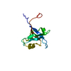
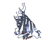
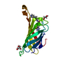
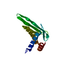
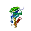
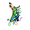
| Title | Anti-Zaire ebolavirus Nucleoprotein Single Domain Antibody Zaire C (ZC) | ||||||||||||
 Components Components | Anti-Zaire ebolavirus Nucleoprotein Single Domain Antibody Zaire C (ZC) | ||||||||||||
 Keywords Keywords | IMMUNE SYSTEM / Antibody / nanobody / Ebola / filovirus | ||||||||||||
| Biological species |  | ||||||||||||
| Method |  X-RAY DIFFRACTION / SYNCHROTRON / MOLECULAR REPLACEMENT / Resolution: 1.49 Å X-RAY DIFFRACTION / SYNCHROTRON / MOLECULAR REPLACEMENT / Resolution: 1.49 Å | ||||||||||||
 Authors Authors | Taylor, A.B. / Sherwood, L.J. / Hart, P.J. / Hayhurst, A. | ||||||||||||
| Funding support |  United States, 3items United States, 3items
| ||||||||||||
 Citation Citation | Journal: J.Mol.Biol. / Year: 2019 Title: Paratope Duality and Gullying are Among the Atypical Recognition Mechanisms Used by a Trio of Nanobodies to Differentiate Ebolavirus Nucleoproteins. Authors: Sherwood, L.J. / Taylor, A.B. / Hart, P.J. / Hayhurst, A. | ||||||||||||
| History |
|
- Structure visualization
| Structure viewer | Molecule:  MolmilJmol/JSmol MolmilJmol/JSmol |
|---|
- Downloads & links
-Download
| PDBx/mmCIF format | 6u53.cif.gz | 69.8 KB | Display | PDBx/mmCIF format |
|---|---|---|---|---|
| PDB format | pdb6u53.ent.gz | 47.8 KB | Display | PDB format |
| PDBx/mmJSON format | 6u53.json.gz | Tree view | PDBx/mmJSON format | |
| Others |  Other downloads Other downloads |
-Validation report
| Arichive directory | https://data.pdbj.org/pub/pdb/validation_reports/u5/6u53ftp://data.pdbj.org/pub/pdb/validation_reports/u5/6u53 | HTTPS FTP |
|---|
-Related structure data
| Related structure data | 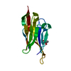 6u50C 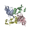 6u51SC 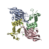 6u52C 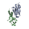 6u54C 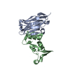 6u55C S: Starting model for refinement C: citing same article ( |
|---|---|
| Similar structure data |


-Links
 PDBj
PDBj


- Assembly
| Deposited unit | 
| ||||||||||||
|---|---|---|---|---|---|---|---|---|---|---|---|---|---|
| 1 |
| ||||||||||||
| Unit cell |
|
-Components
| #1: Antibody | Mass: 13376.943 Da / Num. of mol.: 1 Source method: isolated from a genetically manipulated source Details: Semi-synthetic single pot library Nomad 1 based upon Lama glama Source: (gene. exp.)  |
|---|---|
| #2: Water | ChemComp-HOH /  Mass: 18.015 Da / Num. of mol.: 66 / Source method: isolated from a natural source / Formula: H2O Mass: 18.015 Da / Num. of mol.: 66 / Source method: isolated from a natural source / Formula: H2O |
| Has protein modification | Y |
-Experimental details
-Experiment
| Experiment | Method: X-RAY DIFFRACTION / Number of used crystals: 1 |
|---|
- Sample preparation
| Crystal | Density Matthews: 2.2 Å3/Da / Density % sol: 44.05 % |
|---|---|
| Crystal grow | Temperature: 277 K / Method: vapor diffusion, sitting drop Details: 1.0 M sodium/potassium tartrate, 0.2 M lithium sulfate, 0.1M Tris pH 7.0 |
-Data collection
| Diffraction | Mean temperature: 100 K / Serial crystal experiment: N |
|---|---|
| Diffraction source | Source: SYNCHROTRON / Site: APS / Beamline: 24-ID-C / Wavelength: 0.9791 Å |
| Detector | Type: DECTRIS PILATUS 6M-F / Detector: PIXEL / Date: Apr 8, 2018 |
| Radiation | Protocol: SINGLE WAVELENGTH / Monochromatic (M) / Laue (L): M / Scattering type: x-ray |
| Radiation wavelength | Wavelength: 0.9791 Å / Relative weight: 1 |
| Reflection | Resolution: 1.49→31.24 Å / Num. obs: 13542 / % possible obs: 68.3 % / Redundancy: 12.3 % / Biso Wilson estimate: 29.18 Å2 / CC1/2: 1 / Rpim(I) all: 0.012 / Rrim(I) all: 0.043 / Net I/σ(I): 23.3 |
| Reflection shell | Resolution: 1.49→1.58 Å / Redundancy: 12.3 % / Mean I/σ(I) obs: 1.2 / Num. unique obs: 678 / CC1/2: 0.557 / Rpim(I) all: 0.555 / Rrim(I) all: 1.97 / % possible all: 21.7 |
- Processing
| Software |
| |||||||||||||||||||||||||||||||||||||||||||||||||||||||||||||||||||||||||||||
|---|---|---|---|---|---|---|---|---|---|---|---|---|---|---|---|---|---|---|---|---|---|---|---|---|---|---|---|---|---|---|---|---|---|---|---|---|---|---|---|---|---|---|---|---|---|---|---|---|---|---|---|---|---|---|---|---|---|---|---|---|---|---|---|---|---|---|---|---|---|---|---|---|---|---|---|---|---|---|
| Refinement | Method to determine structure: MOLECULAR REPLACEMENT Starting model: 6U51 Resolution: 1.49→31.24 Å / SU ML: 0.2049 / Cross valid method: THROUGHOUT / σ(F): 1.35 / Phase error: 38.3306
| |||||||||||||||||||||||||||||||||||||||||||||||||||||||||||||||||||||||||||||
| Solvent computation | Shrinkage radii: 0.9 Å / VDW probe radii: 1.11 Å | |||||||||||||||||||||||||||||||||||||||||||||||||||||||||||||||||||||||||||||
| Displacement parameters | Biso mean: 35.87 Å2 | |||||||||||||||||||||||||||||||||||||||||||||||||||||||||||||||||||||||||||||
| Refinement step | Cycle: LAST / Resolution: 1.49→31.24 Å
| |||||||||||||||||||||||||||||||||||||||||||||||||||||||||||||||||||||||||||||
| Refine LS restraints |
| |||||||||||||||||||||||||||||||||||||||||||||||||||||||||||||||||||||||||||||
| LS refinement shell |
|
