- PDB-6u04: Crystal structure of human BRPF1 PZP bound to histone H3 tail -
+
Open data
ID or keywords:
Loading...
-
Basic information
Entry
Database: PDB / ID: 6u04
Title
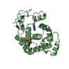
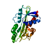
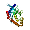
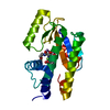
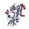
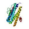
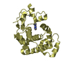
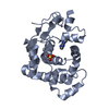
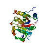
Crystal structure of human BRPF1 PZP bound to histone H3 tail
Components
Histone H3.3,BRPF1, Peregrin
Keywords
TRANSCRIPTION / epigenetics / nucleosome / DNA binding
Function / homology
Function and homology information
Barr body / acetyltransferase activator activity / negative regulation of chromosome condensation / pericentric heterochromatin formation / inner kinetochore / muscle cell differentiation / oocyte maturation / MOZ/MORF histone acetyltransferase complex / regulation of developmental process / regulation of hemopoiesis ...Barr body / acetyltransferase activator activity / negative regulation of chromosome condensation / pericentric heterochromatin formation / inner kinetochore / muscle cell differentiation / oocyte maturation / MOZ/MORF histone acetyltransferase complex / regulation of developmental process / regulation of hemopoiesis / nucleosomal DNA binding / nucleus organization / histone acetyltransferase complex / spermatid development / single fertilization / subtelomeric heterochromatin formation / RNA polymerase II core promoter sequence-specific DNA binding / Regulation of TP53 Activity through Acetylation / Replacement of protamines by nucleosomes in the male pronucleus / embryo implantation / telomere organization / RNA Polymerase I Promoter Opening / Inhibition of DNA recombination at telomere / Assembly of the ORC complex at the origin of replication / Regulation of endogenous retroelements by the Human Silencing Hub (HUSH) complex / DNA methylation / Condensation of Prophase Chromosomes / Chromatin modifications during the maternal to zygotic transition (MZT) / SIRT1 negatively regulates rRNA expression / ERCC6 (CSB) and EHMT2 (G9a) positively regulate rRNA expression / PRC2 methylates histones and DNA / Regulation of endogenous retroelements by KRAB-ZFP proteins / Defective pyroptosis / Regulation of endogenous retroelements by Piwi-interacting RNAs (piRNAs) / RNA Polymerase I Promoter Escape / Transcriptional regulation by small RNAs / Formation of the beta-catenin:TCF transactivating complex / Activated PKN1 stimulates transcription of AR (androgen receptor) regulated genes KLK2 and KLK3 / RUNX1 regulates genes involved in megakaryocyte differentiation and platelet function / NoRC negatively regulates rRNA expression / Negative Regulation of CDH1 Gene Transcription / B-WICH complex positively regulates rRNA expression / male gonad development / Meiotic recombination / Pre-NOTCH Transcription and Translation / multicellular organism growth / Activation of anterior HOX genes in hindbrain development during early embryogenesis / Transcriptional regulation of granulopoiesis / osteoblast differentiation / structural constituent of chromatin / nucleosome / nucleosome assembly / HATs acetylate histones / Factors involved in megakaryocyte development and platelet production / MLL4 and MLL3 complexes regulate expression of PPARG target genes in adipogenesis and hepatic steatosis / RUNX1 regulates transcription of genes involved in differentiation of HSCs / positive regulation of cell growth / Senescence-Associated Secretory Phenotype (SASP) / Oxidative Stress Induced Senescence / Estrogen-dependent gene expression / chromosome, telomeric region / cell population proliferation / RNA polymerase II cis-regulatory region sequence-specific DNA binding / chromatin remodeling / Amyloid fiber formation / protein heterodimerization activity / regulation of transcription by RNA polymerase II / regulation of DNA-templated transcription / positive regulation of DNA-templated transcription / protein-containing complex / DNA binding / extracellular exosome / extracellular region / zinc ion binding / nucleoplasm / nucleus / plasma membrane / cytoplasm / cytosol Similarity search - Function
In the structure databanks used in Yorodumi, some data are registered as the other names, "COVID-19 virus" and "2019-nCoV". Here are the details of the virus and the list of structure data.
Jan 31, 2019. EMDB accession codes are about to change! (news from PDBe EMDB page)
EMDB accession codes are about to change! (news from PDBe EMDB page)
The allocation of 4 digits for EMDB accession codes will soon come to an end. Whilst these codes will remain in use, new EMDB accession codes will include an additional digit and will expand incrementally as the available range of codes is exhausted. The current 4-digit format prefixed with “EMD-” (i.e. EMD-XXXX) will advance to a 5-digit format (i.e. EMD-XXXXX), and so on. It is currently estimated that the 4-digit codes will be depleted around Spring 2019, at which point the 5-digit format will come into force.
The EM Navigator/Yorodumi systems omit the EMD- prefix.
Related info.:Q: What is EMD? / ID/Accession-code notation in Yorodumi/EM Navigator
Yorodumi is a browser for structure data from EMDB, PDB, SASBDB, etc.
This page is also the successor to EM Navigator detail page, and also detail information page/front-end page for Omokage search.
The word "yorodu" (or yorozu) is an old Japanese word meaning "ten thousand". "mi" (miru) is to see.
Related info.:EMDB / PDB / SASBDB / Comparison of 3 databanks / Yorodumi Search / Aug 31, 2016. New EM Navigator & Yorodumi / Yorodumi Papers / Jmol/JSmol / Function and homology information / Changes in new EM Navigator and Yorodumi
 Movie
Movie Controller


 Open data
 Basic information
 Components
 Keywords
 Function and homology information
 Homo sapiens (human)
 X-RAY DIFFRACTION /
  Authors
 Citation
 Structure visualization
 Downloads & links
 Other downloads
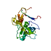

 PDBj
PDBj


 Assembly

 Mass: 140.908 Da / Num. of mol.: 1 / Source method: obtained synthetically / Formula: Pr
 Mass: 65.409 Da / Num. of mol.: 5 / Source method: obtained synthetically / Formula: Zn
 Mass: 18.015 Da / Num. of mol.: 108 / Source method: isolated from a natural source / Formula: H2O
 Sample preparation
 Processing