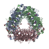
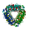
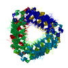
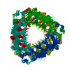
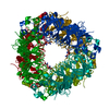
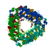
#1: Journal: J Struct Biol / Year: 2021 Title: Practical considerations for using K3 cameras in CDS mode for high-resolution and high-throughput single particle cryo-EM. Authors: Ming Sun / Caleigh M Azumaya / Eric Tse / David P Bulkley / Matthew B Harrington / Glenn Gilbert / Adam Frost / Daniel Southworth / Kliment A Verba / Yifan Cheng / David A Agard / Abstract: Detector technology plays a pivotal role in high-resolution and high-throughput cryo-EM structure determination. Compared with the first-generation, single-electron counting direct detection camera ...Detector technology plays a pivotal role in high-resolution and high-throughput cryo-EM structure determination. Compared with the first-generation, single-electron counting direct detection camera (Gatan K2), the latest K3 camera is faster, larger, and now offers a correlated-double sampling mode (CDS). Importantly this results in a higher DQE and improved throughput compared to its predecessor. In this study, we focused on optimizing camera data collection parameters for daily use within a cryo-EM facility and explored the balance between throughput and resolution. In total, eight data sets of murine heavy-chain apoferritin were collected at different dose rates and magnifications, using 9-hole image shift data collection strategies. The performance of the camera was characterized by the quality of the resultant 3D reconstructions. Our results demonstrated that the Gatan K3 operating in CDS mode outperformed standard (nonCDS) mode in terms of reconstruction resolution in all tested conditions with 8 electrons per pixel per second being the optimal dose rate. At low magnification (64kx) we were able to achieve reconstruction resolutions of 149% of the physical Nyquist limit (1.8 Å with a 1.346 Å physical pixel size). Low magnification allows more particles to be collected per image, aiding analysis of heterogeneous samples requiring large data sets. At moderate magnification (105kx, 0.834 Å physical pixel size) we achieved a resolution of 1.65 Å within 8-h of data collection, a condition optimal for achieving high-resolution on well behaved samples. Our results also show that for an optimal sample like apoferritin, one can achieve better than 2.5 Å resolution with 5 min of data collection. Together, our studies validate the most efficient ways of imaging protein complexes using the K3 direct detector and will greatly benefit the cryo-EM community.
In the structure databanks used in Yorodumi, some data are registered as the other names, "COVID-19 virus" and "2019-nCoV". Here are the details of the virus and the list of structure data.
Jan 31, 2019. EMDB accession codes are about to change! (news from PDBe EMDB page)
EMDB accession codes are about to change! (news from PDBe EMDB page)
The allocation of 4 digits for EMDB accession codes will soon come to an end. Whilst these codes will remain in use, new EMDB accession codes will include an additional digit and will expand incrementally as the available range of codes is exhausted. The current 4-digit format prefixed with “EMD-” (i.e. EMD-XXXX) will advance to a 5-digit format (i.e. EMD-XXXXX), and so on. It is currently estimated that the 4-digit codes will be depleted around Spring 2019, at which point the 5-digit format will come into force.
The EM Navigator/Yorodumi systems omit the EMD- prefix.
Related info.:Q: What is EMD? / ID/Accession-code notation in Yorodumi/EM Navigator
Yorodumi is a browser for structure data from EMDB, PDB, SASBDB, etc.
This page is also the successor to EM Navigator detail page, and also detail information page/front-end page for Omokage search.
The word "yorodu" (or yorozu) is an old Japanese word meaning "ten thousand". "mi" (miru) is to see.
Related info.:EMDB / PDB / SASBDB / Comparison of 3 databanks / Yorodumi Search / Aug 31, 2016. New EM Navigator & Yorodumi / Yorodumi Papers / Jmol/JSmol / Function and homology information / Changes in new EM Navigator and Yorodumi
 Movie
Movie Controller


 Open data
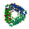
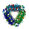
 Basic information
 Components
 Keywords
 Function and homology information
 X-RAY DIFFRACTION /
  Authors
 United States, 1items
  Citation
 Structure visualization
 Downloads & links
 Other downloads
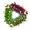


 PDBj
PDBj Assembly
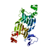

 Mass: 54.938 Da / Num. of mol.: 2 / Source method: obtained synthetically / Formula: Mn
 Mass: 18.015 Da / Num. of mol.: 253 / Source method: isolated from a natural source / Formula: H2O
 Sample preparation
 Processing