 Movie
Movie Controller

[English] 日本語
 Yorodumi
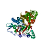
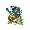
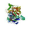
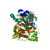
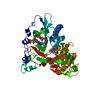
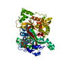
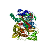
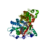
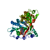
- PDB-6tw7: Leishmania major N-myristoyltransferase in complex with indazole ... -
+ Open data

- Basic information
| Entry | Database: PDB / ID: 6tw7 | ||||||
|---|---|---|---|---|---|---|---|
| Title | Leishmania major N-myristoyltransferase in complex with indazole inhibitor IMP-918 | ||||||
 Components Components | Glycylpeptide N-tetradecanoyltransferase | ||||||
 Keywords Keywords | TRANSFERASE / myristoylation / indazole / Leishmania | ||||||
| Function / homology |  Function and homology information Function and homology informationglycylpeptide N-tetradecanoyltransferase / glycylpeptide N-tetradecanoyltransferase activity / protein localization to membrane / metal ion binding / cytosol Similarity search - Function | ||||||
| Biological species |  Leishmania major (eukaryote) Leishmania major (eukaryote) | ||||||
| Method |  X-RAY DIFFRACTION / SYNCHROTRON / FOURIER SYNTHESIS / Resolution: 1.4 Å X-RAY DIFFRACTION / SYNCHROTRON / FOURIER SYNTHESIS / Resolution: 1.4 Å | ||||||
 Authors Authors | Brannigan, J.A. | ||||||
| Funding support | 1items
| ||||||
 Citation Citation | Journal: To Be Published Title: Leishmania major N-myristoyltransferase in complex with indazole inhibitor IMP-918 Authors: Brannigan, J.A. | ||||||
| History |
|
- Structure visualization
| Structure viewer | Molecule: MolmilJmol/JSmol |
|---|
- Downloads & links
-Download
| PDBx/mmCIF format | 6tw7.cif.gz | 116.4 KB | Display | PDBx/mmCIF format |
|---|---|---|---|---|
| PDB format | pdb6tw7.ent.gz | Display | PDB format | |
| PDBx/mmJSON format | 6tw7.json.gz | Tree view | PDBx/mmJSON format | |
| Others |  Other downloads Other downloads |
-Validation report
| Arichive directory | https://data.pdbj.org/pub/pdb/validation_reports/tw/6tw7ftp://data.pdbj.org/pub/pdb/validation_reports/tw/6tw7 | HTTPS FTP |
|---|
-Related structure data
| Similar structure data |
|---|


-Links
 PDBj
PDBj

- Assembly
| Deposited unit | 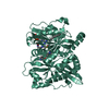
| ||||||||
|---|---|---|---|---|---|---|---|---|---|
| 1 |
| ||||||||
| Unit cell |
|
-Components
| #1: Protein | Mass: 48195.770 Da / Num. of mol.: 1 Source method: isolated from a genetically manipulated source Source: (gene. exp.) Leishmania major (eukaryote) / Gene: NMT, LMJF_32_0080 / Production host:  References: UniProt: Q4Q5S8, glycylpeptide N-tetradecanoyltransferase |
|---|---|
| #2: Chemical | ChemComp-MYA /   Mass: 977.890 Da / Num. of mol.: 1 / Source method: obtained synthetically / Formula: C35H62N7O17P3S Mass: 977.890 Da / Num. of mol.: 1 / Source method: obtained synthetically / Formula: C35H62N7O17P3S |
| #3: Chemical | ChemComp-MG /   Mass: 24.305 Da / Num. of mol.: 1 / Source method: obtained synthetically / Formula: Mg Mass: 24.305 Da / Num. of mol.: 1 / Source method: obtained synthetically / Formula: Mg |
| #4: Chemical | ChemComp-NZB / [(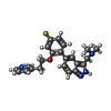  Mass: 390.453 Da / Num. of mol.: 1 / Source method: obtained synthetically / Formula: C23H23FN4O / Feature type: SUBJECT OF INVESTIGATION Mass: 390.453 Da / Num. of mol.: 1 / Source method: obtained synthetically / Formula: C23H23FN4O / Feature type: SUBJECT OF INVESTIGATION |
| #5: Water | ChemComp-HOH /  Mass: 18.015 Da / Num. of mol.: 406 / Source method: isolated from a natural source / Formula: H2O Mass: 18.015 Da / Num. of mol.: 406 / Source method: isolated from a natural source / Formula: H2O |
| Has ligand of interest | Y |
-Experimental details
-Experiment
| Experiment | Method: X-RAY DIFFRACTION / Number of used crystals: 1 |
|---|
- Sample preparation
| Crystal | Density Matthews: 2.23 Å3/Da / Density % sol: 45 % |
|---|---|
| Crystal grow | Temperature: 291 K / Method: vapor diffusion, sitting drop / pH: 5.5 Details: 30% PEG 1500, 0.2 M NaCl, 0.1 M Na cacodylate, pH 5.5 |
-Data collection
| Diffraction | Mean temperature: 100 K / Serial crystal experiment: N |
|---|---|
| Diffraction source | Source: SYNCHROTRON / Site: Diamond  / Beamline: I03 / Wavelength: 0.9763 Å / Beamline: I03 / Wavelength: 0.9763 Å |
| Detector | Type: DECTRIS PILATUS 2M / Detector: PIXEL / Date: May 2, 2013 |
| Radiation | Protocol: SINGLE WAVELENGTH / Monochromatic (M) / Laue (L): M / Scattering type: x-ray |
| Radiation wavelength | Wavelength: 0.9763 Å / Relative weight: 1 |
| Reflection | Resolution: 1.4→49 Å / Num. obs: 83675 / % possible obs: 99.3 % / Redundancy: 4 % / Rmerge(I) obs: 0.034 / Net I/σ(I): 14.8 |
| Reflection shell | Resolution: 1.4→1.42 Å / Redundancy: 2.8 % / Rmerge(I) obs: 0.802 / Mean I/σ(I) obs: 1.3 / Num. unique obs: 3809 / % possible all: 91.6 |
- Processing
| Software |
| |||||||||||||||||||||||||||||||||||||||||||||||||||||||||||||||||||||||||||||||||||||||||||||||||||||||||||||||||||||||||||||||||||||||||||||||||||||||||||||||||||||||||||||||||||||||||||||||||||||||||||||||||||||||||||||||||||||||
|---|---|---|---|---|---|---|---|---|---|---|---|---|---|---|---|---|---|---|---|---|---|---|---|---|---|---|---|---|---|---|---|---|---|---|---|---|---|---|---|---|---|---|---|---|---|---|---|---|---|---|---|---|---|---|---|---|---|---|---|---|---|---|---|---|---|---|---|---|---|---|---|---|---|---|---|---|---|---|---|---|---|---|---|---|---|---|---|---|---|---|---|---|---|---|---|---|---|---|---|---|---|---|---|---|---|---|---|---|---|---|---|---|---|---|---|---|---|---|---|---|---|---|---|---|---|---|---|---|---|---|---|---|---|---|---|---|---|---|---|---|---|---|---|---|---|---|---|---|---|---|---|---|---|---|---|---|---|---|---|---|---|---|---|---|---|---|---|---|---|---|---|---|---|---|---|---|---|---|---|---|---|---|---|---|---|---|---|---|---|---|---|---|---|---|---|---|---|---|---|---|---|---|---|---|---|---|---|---|---|---|---|---|---|---|---|---|---|---|---|---|---|---|---|---|---|---|---|---|---|---|---|---|
| Refinement | Method to determine structure: FOURIER SYNTHESIS / Resolution: 1.4→49 Å / Cor.coef. Fo:Fc: 0.968 / Cor.coef. Fo:Fc free: 0.955 / Cross valid method: FREE R-VALUE / ESU R: 0.073 / ESU R Free: 0.078 Details: Hydrogens have been added in their riding positions
| |||||||||||||||||||||||||||||||||||||||||||||||||||||||||||||||||||||||||||||||||||||||||||||||||||||||||||||||||||||||||||||||||||||||||||||||||||||||||||||||||||||||||||||||||||||||||||||||||||||||||||||||||||||||||||||||||||||||
| Solvent computation | Ion probe radii: 0.8 Å / Shrinkage radii: 0.8 Å / VDW probe radii: 1.2 Å | |||||||||||||||||||||||||||||||||||||||||||||||||||||||||||||||||||||||||||||||||||||||||||||||||||||||||||||||||||||||||||||||||||||||||||||||||||||||||||||||||||||||||||||||||||||||||||||||||||||||||||||||||||||||||||||||||||||||
| Displacement parameters | Biso mean: 25.298 Å2
| |||||||||||||||||||||||||||||||||||||||||||||||||||||||||||||||||||||||||||||||||||||||||||||||||||||||||||||||||||||||||||||||||||||||||||||||||||||||||||||||||||||||||||||||||||||||||||||||||||||||||||||||||||||||||||||||||||||||
| Refinement step | Cycle: LAST / Resolution: 1.4→49 Å
| |||||||||||||||||||||||||||||||||||||||||||||||||||||||||||||||||||||||||||||||||||||||||||||||||||||||||||||||||||||||||||||||||||||||||||||||||||||||||||||||||||||||||||||||||||||||||||||||||||||||||||||||||||||||||||||||||||||||
| Refine LS restraints |
| |||||||||||||||||||||||||||||||||||||||||||||||||||||||||||||||||||||||||||||||||||||||||||||||||||||||||||||||||||||||||||||||||||||||||||||||||||||||||||||||||||||||||||||||||||||||||||||||||||||||||||||||||||||||||||||||||||||||
| LS refinement shell | Refine-ID: X-RAY DIFFRACTION / Total num. of bins used: 20
|
