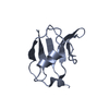
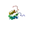
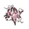
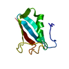
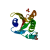
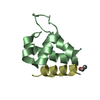
[日本語] English
- PDB-6ts3: EF-hands 3 and 4 of alpha-actinin in complex with CaMKII regulato... -

+
データを開く


IDまたはキーワード:

読み込み中...

-
基本情報

登録情報
データベース: PDB / ID: 6ts3
タイトルEF-hands 3 and 4 of alpha-actinin in complex with CaMKII regulatory segment
要素
  • ACE-ASN-ALA-ARG-ARG-LYS-LEU-LYS-GLY-ALA-ILE-LEU-THR-THR-MET-LEU-ALA-THR-ARG-ASN-PHE
  • Alpha-actinin-2
キーワードPEPTIDE BINDING PROTEIN / CaMKII-binding protein
機能・相同性
機能・相同性情報


regulation of synaptic vesicle docking / actin filament uncapping / FATZ binding / titin Z domain binding / HSF1-dependent transactivation / Interferon gamma signaling / positive regulation of potassium ion transmembrane transporter activity / phospholipase C-activating angiotensin-activated signaling pathway / positive regulation of endocytic recycling / Ion transport by P-type ATPases ...regulation of synaptic vesicle docking / actin filament uncapping / FATZ binding / titin Z domain binding / HSF1-dependent transactivation / Interferon gamma signaling / positive regulation of potassium ion transmembrane transporter activity / phospholipase C-activating angiotensin-activated signaling pathway / positive regulation of endocytic recycling / Ion transport by P-type ATPases / peptidyl-threonine autophosphorylation / Unblocking of NMDA receptors, glutamate binding and activation / regulation of endocannabinoid signaling pathway / RAF activation / calcium- and calmodulin-dependent protein kinase complex / Trafficking of AMPA receptors / Ca2+ pathway / RAF/MAP kinase cascade / Ca2+/calmodulin-dependent protein kinase / negative regulation of potassium ion transmembrane transporter activity / negative regulation of protein localization to cell surface / positive regulation of cation channel activity / LIM domain binding / microspike assembly / negative regulation of hydrolase activity / dendritic spine development / regulation of neuron migration / regulation of neurotransmitter secretion / postsynaptic actin cytoskeleton / structural constituent of postsynaptic actin cytoskeleton / positive regulation of potassium ion transport / Ion homeostasis / focal adhesion assembly / muscle cell development / positive regulation of calcium ion transport / Striated Muscle Contraction / calcium/calmodulin-dependent protein kinase activity / Nephrin family interactions / Assembly and cell surface presentation of NMDA receptors / regulation of mitochondrial membrane permeability involved in apoptotic process / cardiac muscle cell development / structural constituent of muscle / dendrite morphogenesis / sarcomere organization / cortical actin cytoskeleton / Negative regulation of NMDA receptor-mediated neuronal transmission / Unblocking of NMDA receptors, glutamate binding and activation / pseudopodium / negative regulation of potassium ion transport / regulation of neuronal synaptic plasticity / Long-term potentiation / postsynaptic density, intracellular component / glutamate receptor binding / cellular response to interferon-beta / positive regulation of cardiac muscle cell apoptotic process / cytoskeletal protein binding / titin binding / phosphatidylinositol-4,5-bisphosphate binding / Ras activation upon Ca2+ influx through NMDA receptor / platelet alpha granule lumen / response to ischemia / protein localization to plasma membrane / regulation of membrane potential / cell projection / positive regulation of receptor signaling pathway via JAK-STAT / filopodium / actin filament / postsynaptic density membrane / Schaffer collateral - CA1 synapse / Z disc / G1/S transition of mitotic cell cycle / calcium ion transport / actin filament binding / integrin binding / cell junction / Platelet degranulation / actin cytoskeleton organization / RAF/MAP kinase cascade / regulation of apoptotic process / dendritic spine / transmembrane transporter binding / cytoskeleton / calmodulin binding / postsynaptic density / cell adhesion / protein domain specific binding / protein serine kinase activity / focal adhesion / protein serine/threonine kinase activity / synapse / calcium ion binding / glutamatergic synapse / mitochondrion / extracellular exosome / extracellular region / ATP binding / metal ion binding / identical protein binding / cytosol
類似検索 - 分子機能
Calcium/calmodulin-dependent protein kinase II, association-domain / Calcium/calmodulin dependent protein kinase II association domain / EF-hand, Ca insensitive / Ca2+ insensitive EF hand / Ca2+ insensitive EF hand / Spectrin repeat / Spectrin repeat / Spectrin/alpha-actinin / Spectrin repeats / Actinin-type actin-binding domain signature 1. ...Calcium/calmodulin-dependent protein kinase II, association-domain / Calcium/calmodulin dependent protein kinase II association domain / EF-hand, Ca insensitive / Ca2+ insensitive EF hand / Ca2+ insensitive EF hand / Spectrin repeat / Spectrin repeat / Spectrin/alpha-actinin / Spectrin repeats / Actinin-type actin-binding domain signature 1. / Actinin-type actin-binding domain signature 2. / Actinin-type actin-binding domain, conserved site / Calponin homology domain / Calponin homology (CH) domain / Calponin homology domain / CH domain superfamily / Calponin homology (CH) domain profile. / NTF2-like domain superfamily / EF-hand domain pair / EF-hand, calcium binding motif / EF-hand calcium-binding domain profile. / EF-hand domain / EF-hand domain pair / Serine/threonine-protein kinase, active site / Serine/Threonine protein kinases active-site signature. / Protein kinase domain / Serine/Threonine protein kinases, catalytic domain / Protein kinase, ATP binding site / Protein kinases ATP-binding region signature. / Protein kinase domain profile. / Protein kinase domain / Protein kinase-like domain superfamily
類似検索 - ドメイン・相同性
ACETYL GROUP / Calcium/calmodulin-dependent protein kinase type II subunit alpha / Alpha-actinin-2
類似検索 - 構成要素
生物種Homo sapiens (ヒト)
Mus musculus (ハツカネズミ)
手法X線回折 / シンクロトロン / 単波長異常分散 / 解像度: 1.28 Å
データ登録者Zhu, J. / Gold, M.
資金援助 英国, 1件
組織認可番号
Biotechnology and Biological Sciences Research Council (BBSRC)533303 英国
引用ジャーナル: To Be Published
タイトル: EF-hands 3 and 4 of alpha-actinin in complex with CaMKII regulatory segment
著者: Zhu, J. / Gold, M.
履歴
登録2019年12月19日登録サイト: PDBE / 処理サイト: PDBE
改定 1.02021年1月13日Provider: repository / タイプ: Initial release
改定 1.12025年4月9日Group: Data collection / Database references / Structure summary
カテゴリ: chem_comp_atom / chem_comp_bond ...chem_comp_atom / chem_comp_bond / database_2 / pdbx_entry_details
Item: _database_2.pdbx_DOI / _database_2.pdbx_database_accession / _pdbx_entry_details.has_protein_modification

-
構造の表示

構造ビューア分子:
MolmilJmol/JSmol

ダウンロードとリンク

-
集合体

登録構造単位
A: Alpha-actinin-2
B: Alpha-actinin-2
C: ACE-ASN-ALA-ARG-ARG-LYS-LEU-LYS-GLY-ALA-ILE-LEU-THR-THR-MET-LEU-ALA-THR-ARG-ASN-PHE
D: ACE-ASN-ALA-ARG-ARG-LYS-LEU-LYS-GLY-ALA-ILE-LEU-THR-THR-MET-LEU-ALA-THR-ARG-ASN-PHE
ヘテロ分子


分子量 (理論値)分子数
合計 (水以外)20,7456
ポリマ-20,6574
非ポリマー882
4,792266
1
A: Alpha-actinin-2
C: ACE-ASN-ALA-ARG-ARG-LYS-LEU-LYS-GLY-ALA-ILE-LEU-THR-THR-MET-LEU-ALA-THR-ARG-ASN-PHE
ヘテロ分子


分子量 (理論値)分子数
合計 (水以外)10,3733
ポリマ-10,3292
非ポリマー441
362
タイプ名称対称操作
identity operation1_555x,y,z1
Buried area1510 Å2
ΔGint-9 kcal/mol
Surface area5640 Å2
手法PISA
2
B: Alpha-actinin-2
D: ACE-ASN-ALA-ARG-ARG-LYS-LEU-LYS-GLY-ALA-ILE-LEU-THR-THR-MET-LEU-ALA-THR-ARG-ASN-PHE
ヘテロ分子


分子量 (理論値)分子数
合計 (水以外)10,3733
ポリマ-10,3292
非ポリマー441
362
タイプ名称対称操作
identity operation1_555x,y,z1
Buried area1240 Å2
ΔGint-9 kcal/mol
Surface area5110 Å2
手法PISA
単位格子
Length a, b, c (Å)43.355, 47.270, 47.472
Angle α, β, γ (deg.)90.000, 110.532, 90.000
Int Tables number4
Space group name H-MP1211
Space group name HallP2yb
Symmetry operation#1: x,y,z
#2: -x,y+1/2,-z

-
要素

#1: タンパク質 Alpha-actinin-2 / Alpha-actinin skeletal muscle isoform 2 / F-actin cross-linking protein


分子量: 7903.778 Da / 分子数: 2 / 由来タイプ: 組換発現 / 由来: (組換発現) Homo sapiens (ヒト) / 遺伝子: ACTN2 / 発現宿主: Escherichia coli (大腸菌) / 参照: UniProt: P35609
#2: タンパク質・ペプチド ACE-ASN-ALA-ARG-ARG-LYS-LEU-LYS-GLY-ALA-ILE-LEU-THR-THR-MET-LEU-ALA-THR-ARG-ASN-PHE


分子量: 2424.889 Da / 分子数: 2 / 由来タイプ: 合成 / 由来: (合成) Mus musculus (ハツカネズミ) / 参照: UniProt: P11798*PLUS
#3: 化合物 ChemComp-ACE / ACETYL GROUP / アセトアルデヒド


分子量: 44.053 Da / 分子数: 2 / 由来タイプ: 合成 / : C2H4O / タイプ: SUBJECT OF INVESTIGATION
#4: 水 ChemComp-HOH / water


分子量: 18.015 Da / 分子数: 266 / 由来タイプ: 天然 / : H2O
研究の焦点であるリガンドがあるかY
Has protein modificationN

-
実験情報

-
実験

実験手法: X線回折 / 使用した結晶の数: 1

-
試料調製

結晶マシュー密度: 2.2 Å3/Da / 溶媒含有率: 44.15 %
結晶化温度: 277 K / 手法: 蒸気拡散法, シッティングドロップ法
詳細: 0.1 M BIS-TRIS pH 6.5, 25% w/v Polyethylene glycol 3,350

-
データ収集

回折平均測定温度: 100 K / Serial crystal experiment: N
放射光源由来: シンクロトロン / サイト: Diamond / ビームライン: I04 / 波長: 0.9795 Å
検出器タイプ: DECTRIS EIGER X 16M / 検出器: PIXEL / 日付: 2019年10月14日
放射プロトコル: SINGLE WAVELENGTH / 単色(M)・ラウエ(L): M / 散乱光タイプ: x-ray
放射波長波長: 0.9795 Å / 相対比: 1
反射解像度: 1.28→44.46 Å / Num. obs: 46303 / % possible obs: 99.8 % / 冗長度: 6.6 % / Biso Wilson estimate: 16.75 Å2 / CC1/2: 0.999 / Rmerge(I) obs: 0.035 / Rrim(I) all: 0.038 / Net I/σ(I): 19.7
反射 シェル解像度: 1.28→1.3 Å / Rmerge(I) obs: 0.437 / Mean I/σ(I) obs: 2.4 / Num. unique obs: 2152 / CC1/2: 0.94 / Rrim(I) all: 0.487

-
解析

ソフトウェア
名称バージョン分類
DIALS1.17.1_3660データ収集
PHENIX1.17.1_3660精密化
DIALSデータ削減
Aimlessデータスケーリング
CRANK2位相決定
精密化構造決定の手法: 単波長異常分散 / 解像度: 1.28→44.46 Å / SU ML: 0.1249 / 交差検証法: FREE R-VALUE / σ(F): 1.37 / 位相誤差: 22.6175
Rfactor反射数%反射
Rfree0.1989 1996 4.32 %
Rwork0.1832 --
obs0.1839 46220 99.61 %
溶媒の処理減衰半径: 0.9 Å / VDWプローブ半径: 1.11 Å
原子変位パラメータBiso mean: 25.69 Å2
精密化ステップサイクル: LAST / 解像度: 1.28→44.46 Å
タンパク質核酸リガンド溶媒全体
原子数1352 0 0 266 1618
拘束条件
Refine-IDタイプDev ideal
X-RAY DIFFRACTIONf_bond_d0.00511431
X-RAY DIFFRACTIONf_angle_d0.69821949
X-RAY DIFFRACTIONf_chiral_restr0.0699222
X-RAY DIFFRACTIONf_plane_restr0.005257
X-RAY DIFFRACTIONf_dihedral_angle_d13.0746214
LS精密化 シェル
解像度 (Å)Rfactor RfreeNum. reflection RfreeRfactor RworkNum. reflection RworkRefine-ID% reflection obs (%)
1.28-1.310.29581380.2523044X-RAY DIFFRACTION96.69
1.31-1.350.29071390.24273119X-RAY DIFFRACTION99.76
1.35-1.390.27231420.22973149X-RAY DIFFRACTION99.67
1.39-1.430.2451440.21273167X-RAY DIFFRACTION99.85
1.43-1.480.22141420.20523145X-RAY DIFFRACTION99.58
1.48-1.540.22621420.19783146X-RAY DIFFRACTION99.82
1.54-1.610.20031420.18813147X-RAY DIFFRACTION99.85
1.61-1.70.22291430.18883165X-RAY DIFFRACTION99.97
1.7-1.80.19321430.19453178X-RAY DIFFRACTION99.94
1.8-1.940.19821430.18963145X-RAY DIFFRACTION99.94
1.94-2.140.19111440.18063187X-RAY DIFFRACTION99.97
2.14-2.450.17451430.18193179X-RAY DIFFRACTION100
2.45-3.080.20841460.18593213X-RAY DIFFRACTION99.97
3.09-44.460.18481450.16573240X-RAY DIFFRACTION99.65
精密化 TLS手法: refined / Origin x: 15.1281362844 Å / Origin y: 21.9163990434 Å / Origin z: 51.1831683785 Å
111213212223313233
T0.121159552278 Å20.00884287218521 Å2-0.0138581632918 Å2-0.119212347253 Å20.0120755410377 Å2--0.156698327561 Å2
L1.04206130367 °2-0.438679642654 °2-0.297515697624 °2-0.485852111388 °2-0.0990778256617 °2--2.13401916637 °2
S0.0436904734155 Å °-0.0496646452414 Å °-0.0649457729327 Å °-0.00654105398861 Å °0.0328064869885 Å °0.0391085940183 Å °-0.0135063513924 Å °-0.289520261344 Å °-0.0432803874475 Å °
精密化 TLSグループSelection details: all

+
万見について

-
お知らせ

-
2022年2月9日: EMDBエントリの付随情報ファイルのフォーマットが新しくなりました

EMDBエントリの付随情報ファイルのフォーマットが新しくなりました

  • EMDBのヘッダファイルのバージョン3が、公式のフォーマットとなりました。
  • これまでは公式だったバージョン1.9は、アーカイブから削除されます。

関連情報:EMDBヘッダ

外部リンク:wwPDBはEMDBデータモデルのバージョン3へ移行します

-
2020年8月12日: 新型コロナ情報

新型コロナ情報

URL: https://pdbj.org/emnavi/covid19.php

新ページ: EM Navigatorに新型コロナウイルスの特設ページを開設しました。

関連情報:Covid-19情報 / 2020年3月5日: 新型コロナウイルスの構造データ

+
2020年3月5日: 新型コロナウイルスの構造データ

新型コロナウイルスの構造データ

関連情報:万見生物種 / 2020年8月12日: 新型コロナ情報

外部リンク:COVID-19特集ページ - PDBj / 今月の分子2020年2月:コロナウイルスプロテーアーゼ

+
2019年1月31日: EMDBのIDの桁数の変更

EMDBのIDの桁数の変更

  • EMDBエントリに付与されているアクセスコード(EMDB-ID)は4桁の数字(例、EMD-1234)でしたが、間もなく枯渇します。これまでの4桁のID番号は4桁のまま変更されませんが、4桁の数字を使い切った後に発行されるIDは5桁以上の数字(例、EMD-12345)になります。5桁のIDは2019年の春頃から発行される見通しです。
  • EM Navigator/万見では、接頭語「EMD-」は省略されています。

関連情報:Q: 「EMD」とは何ですか? / 万見/EM NavigatorにおけるID/アクセスコードの表記

外部リンク:EMDB Accession Codes are Changing Soon! / PDBjへお問い合わせ

+
2017年7月12日: PDB大規模アップデート

PDB大規模アップデート

  • 新バージョンのPDBx/mmCIF辞書形式に基づくデータがリリースされました。
  • 今回の更新はバージョン番号が4から5になる大規模なもので、全エントリデータの書き換えが行われる「Remediation」というアップデートに該当します。
  • このバージョンアップで、電子顕微鏡の実験手法に関する多くの項目の書式が改定されました(例:em_softwareなど)。
  • EM NavigatorとYorodumiでも、この改定に基づいた表示内容になります。

外部リンク:wwPDB Remediation / OneDepデータ基準に準拠した、より強化された内容のモデル構造ファイルが、PDBアーカイブで公開されました。

-
万見 (Yorodumi)

幾万の構造データを、幾万の視点から

  • 万見(Yorodumi)は、EMDB/PDB/SASBDBなどの構造データを閲覧するためのページです。
  • EM Navigatorの詳細ページの後継、Omokage検索のフロントエンドも兼ねています。

関連情報:EMDB / PDB / SASBDB / 3つのデータバンクの比較 / 万見検索 / 2016年8月31日: 新しいEM Navigatorと万見 / 万見文献 / Jmol/JSmol / 機能・相同性情報 / 新しいEM Navigatorと万見の変更点

他の情報も見る