 Movie
Movie Controller

[English] 日本語
 Yorodumi
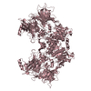
- PDB-6trw: Crystal structure of DPP8 in complex with the EIL peptide (SLRFLF... -
+ Open data

- Basic information
| Entry | Database: PDB / ID: 6trw | ||||||
|---|---|---|---|---|---|---|---|
| Title | Crystal structure of DPP8 in complex with the EIL peptide (SLRFLFEGQRIADNH) | ||||||
 Components Components |
| ||||||
 Keywords Keywords | HYDROLASE / EIL peptide / DPP8 / SUMO1 | ||||||
| Function / homology |  Function and homology information Function and homology informationprotein localization to nuclear pore / SUMOylation of nuclear envelope proteins / SUMO is proteolytically processed / Negative regulation of activity of TFAP2 (AP-2) family transcription factors / SUMO is conjugated to E1 (UBA2:SAE1) / SUMO is transferred from E1 to E2 (UBE2I, UBC9) / negative regulation of transcription initiation by RNA polymerase II / negative regulation of action potential / nuclear stress granule / PML body organization ...protein localization to nuclear pore / SUMOylation of nuclear envelope proteins / SUMO is proteolytically processed / Negative regulation of activity of TFAP2 (AP-2) family transcription factors / SUMO is conjugated to E1 (UBA2:SAE1) / SUMO is transferred from E1 to E2 (UBE2I, UBC9) / negative regulation of transcription initiation by RNA polymerase II / negative regulation of action potential / nuclear stress granule / PML body organization / dipeptidyl-peptidase IV / small protein activating enzyme binding / SUMOylation of immune response proteins / SUMOylation of SUMOylation proteins / regulation of calcium ion transmembrane transport / SUMOylation of DNA methylation proteins / Maturation of nucleoprotein / dipeptidyl-peptidase activity / XY body / SUMOylation of RNA binding proteins / regulation of cardiac muscle cell contraction / Postmitotic nuclear pore complex (NPC) reformation / Maturation of nucleoprotein / negative regulation of protein import into nucleus / negative regulation of programmed cell death / SUMOylation of ubiquitinylation proteins / ubiquitin-specific protease binding / cellular response to cadmium ion / SUMOylation of transcription factors / roof of mouth development / SUMOylation of DNA replication proteins / ubiquitin-like protein ligase binding / protein sumoylation / potassium channel regulator activity / aminopeptidase activity / Regulation of IFNG signaling / nuclear pore / transporter activator activity / postsynaptic cytosol / SUMOylation of DNA damage response and repair proteins / presynaptic cytosol / Transcriptional and post-translational regulation of MITF-M expression and activity / SUMOylation of transcription cofactors / serine-type peptidase activity / SUMOylation of chromatin organization proteins / SUMOylation of intracellular receptors / regulation of protein stability / positive regulation of protein-containing complex assembly / PML body / PKR-mediated signaling / Formation of Incision Complex in GG-NER / protein tag activity / positive regulation of proteasomal ubiquitin-dependent protein catabolic process / cellular response to heat / Recruitment and ATM-mediated phosphorylation of repair and signaling proteins at DNA double strand breaks / nuclear membrane / nuclear speck / protein stabilization / nuclear body / immune response / DNA repair / negative regulation of DNA-templated transcription / apoptotic process / ubiquitin protein ligase binding / nucleolus / glutamatergic synapse / enzyme binding / negative regulation of transcription by RNA polymerase II / proteolysis / RNA binding / nucleoplasm / nucleus / plasma membrane / cytoplasm / cytosol Similarity search - Function | ||||||
| Biological species |  Homo sapiens (human) Homo sapiens (human) | ||||||
| Method |  X-RAY DIFFRACTION / SYNCHROTRON / MOLECULAR REPLACEMENT / Resolution: 3 Å X-RAY DIFFRACTION / SYNCHROTRON / MOLECULAR REPLACEMENT / Resolution: 3 Å | ||||||
 Authors Authors | Ross, B. / Huber, R. | ||||||
 Citation Citation | Journal: J.Appl.Crystallogr. / Year: 2021 Title: Aerosol-based ligand soaking of reservoir-free protein crystals. Authors: Ross, B. / Krapp, S. / Geiss-Friedlander, R. / Littmann, W. / Huber, R. / Kiefersauer, R. | ||||||
| History |
|
- Structure visualization
| Structure viewer | Molecule: MolmilJmol/JSmol |
|---|
- Downloads & links
-Download
| PDBx/mmCIF format | 6trw.cif.gz | 521 KB | Display | PDBx/mmCIF format |
|---|---|---|---|---|
| PDB format | pdb6trw.ent.gz | 420.1 KB | Display | PDB format |
| PDBx/mmJSON format | 6trw.json.gz | Tree view | PDBx/mmJSON format | |
| Others |  Other downloads Other downloads |
-Validation report
| Arichive directory | https://data.pdbj.org/pub/pdb/validation_reports/tr/6trwftp://data.pdbj.org/pub/pdb/validation_reports/tr/6trw | HTTPS FTP |
|---|
-Related structure data
| Related structure data |  6trxC 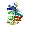 7nvqC  6eooS S: Starting model for refinement C: citing same article ( |
|---|---|
| Similar structure data |


-Links
 PDBj
PDBj


- Assembly
| Deposited unit | 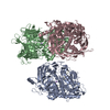
| ||||||||
|---|---|---|---|---|---|---|---|---|---|
| 1 | 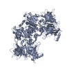
| ||||||||
| 2 | 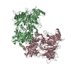
| ||||||||
| Unit cell |
|
-Components
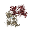
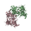
| #1: Protein | Mass: 103886.812 Da / Num. of mol.: 3 Source method: isolated from a genetically manipulated source Source: (gene. exp.) Homo sapiens (human) / Gene: DPP8, DPRP1, MSTP097, MSTP135, MSTP141 / Production host:   Spodoptera frugiperda (fall armyworm) / References: UniProt: Q6V1X1, dipeptidyl-peptidase IV Spodoptera frugiperda (fall armyworm) / References: UniProt: Q6V1X1, dipeptidyl-peptidase IV#2: Protein/peptide | Mass: 1806.011 Da / Num. of mol.: 3 / Source method: obtained synthetically / Source: (synth.) Homo sapiens (human) / References: UniProt: P63165*PLUS#3: Chemical |   Mass: 22.990 Da / Num. of mol.: 3 / Source method: obtained synthetically / Formula: Na Mass: 22.990 Da / Num. of mol.: 3 / Source method: obtained synthetically / Formula: Na#4: Water | ChemComp-HOH / |  Mass: 18.015 Da / Num. of mol.: 33 / Source method: isolated from a natural source / Formula: H2O Mass: 18.015 Da / Num. of mol.: 33 / Source method: isolated from a natural source / Formula: H2OHas ligand of interest | N | |
|---|
-Experimental details
-Experiment
| Experiment | Method: X-RAY DIFFRACTION / Number of used crystals: 1 |
|---|
- Sample preparation
| Crystal | Density Matthews: 4.12 Å3/Da / Density % sol: 70.16 % |
|---|---|
| Crystal grow | Temperature: 277 K / Method: vapor diffusion, hanging drop / pH: 6.75 / Details: 0.46 M Na citrate pH 6.75 |
-Data collection
| Diffraction | Mean temperature: 100 K / Serial crystal experiment: N | ||||||||||||||||||||||||||||||||||||||||||||||||||||||||||||||||||||||||||||||||||||||||||||||||||||||||||||||||||||||||||||||||||||||||||||||||||||||||||||||||||||||||||||||||||||||||||||||
|---|---|---|---|---|---|---|---|---|---|---|---|---|---|---|---|---|---|---|---|---|---|---|---|---|---|---|---|---|---|---|---|---|---|---|---|---|---|---|---|---|---|---|---|---|---|---|---|---|---|---|---|---|---|---|---|---|---|---|---|---|---|---|---|---|---|---|---|---|---|---|---|---|---|---|---|---|---|---|---|---|---|---|---|---|---|---|---|---|---|---|---|---|---|---|---|---|---|---|---|---|---|---|---|---|---|---|---|---|---|---|---|---|---|---|---|---|---|---|---|---|---|---|---|---|---|---|---|---|---|---|---|---|---|---|---|---|---|---|---|---|---|---|---|---|---|---|---|---|---|---|---|---|---|---|---|---|---|---|---|---|---|---|---|---|---|---|---|---|---|---|---|---|---|---|---|---|---|---|---|---|---|---|---|---|---|---|---|---|---|---|---|
| Diffraction source | Source: SYNCHROTRON / Site: SLS  / Beamline: X10SA / Wavelength: 0.99989 Å / Beamline: X10SA / Wavelength: 0.99989 Å | ||||||||||||||||||||||||||||||||||||||||||||||||||||||||||||||||||||||||||||||||||||||||||||||||||||||||||||||||||||||||||||||||||||||||||||||||||||||||||||||||||||||||||||||||||||||||||||||
| Detector | Type: DECTRIS PILATUS 6M-F / Detector: PIXEL / Date: Feb 13, 2019 | ||||||||||||||||||||||||||||||||||||||||||||||||||||||||||||||||||||||||||||||||||||||||||||||||||||||||||||||||||||||||||||||||||||||||||||||||||||||||||||||||||||||||||||||||||||||||||||||
| Radiation | Protocol: SINGLE WAVELENGTH / Monochromatic (M) / Laue (L): M / Scattering type: x-ray | ||||||||||||||||||||||||||||||||||||||||||||||||||||||||||||||||||||||||||||||||||||||||||||||||||||||||||||||||||||||||||||||||||||||||||||||||||||||||||||||||||||||||||||||||||||||||||||||
| Radiation wavelength | Wavelength: 0.99989 Å / Relative weight: 1 | ||||||||||||||||||||||||||||||||||||||||||||||||||||||||||||||||||||||||||||||||||||||||||||||||||||||||||||||||||||||||||||||||||||||||||||||||||||||||||||||||||||||||||||||||||||||||||||||
| Reflection | Resolution: 3→49.17 Å / Num. obs: 104482 / % possible obs: 99.9 % / Redundancy: 8.427 % / Biso Wilson estimate: 77.091 Å2 / CC1/2: 0.999 / Rmerge(I) obs: 0.091 / Rrim(I) all: 0.097 / Χ2: 0.997 / Net I/σ(I): 20.41 / Num. measured all: 880465 / Scaling rejects: 90 | ||||||||||||||||||||||||||||||||||||||||||||||||||||||||||||||||||||||||||||||||||||||||||||||||||||||||||||||||||||||||||||||||||||||||||||||||||||||||||||||||||||||||||||||||||||||||||||||
| Reflection shell | Diffraction-ID: 1
|
- Processing
| Software |
| ||||||||||||||||||||||||||||||||||||||||||||||||||||||||||||
|---|---|---|---|---|---|---|---|---|---|---|---|---|---|---|---|---|---|---|---|---|---|---|---|---|---|---|---|---|---|---|---|---|---|---|---|---|---|---|---|---|---|---|---|---|---|---|---|---|---|---|---|---|---|---|---|---|---|---|---|---|---|
| Refinement | Method to determine structure: MOLECULAR REPLACEMENT Starting model: 6EOO Resolution: 3→49.17 Å / Cor.coef. Fo:Fc: 0.952 / Cor.coef. Fo:Fc free: 0.928 / SU B: 15.252 / SU ML: 0.262 / Cross valid method: THROUGHOUT / σ(F): 0 / ESU R: 0.658 / ESU R Free: 0.316 Details: HYDROGENS HAVE BEEN ADDED IN THE RIDING POSITIONS U VALUES : REFINED INDIVIDUALLY
| ||||||||||||||||||||||||||||||||||||||||||||||||||||||||||||
| Solvent computation | Ion probe radii: 0.8 Å / Shrinkage radii: 0.8 Å / VDW probe radii: 1.2 Å | ||||||||||||||||||||||||||||||||||||||||||||||||||||||||||||
| Displacement parameters | Biso max: 224.53 Å2 / Biso mean: 91.044 Å2 / Biso min: 48.44 Å2
| ||||||||||||||||||||||||||||||||||||||||||||||||||||||||||||
| Refinement step | Cycle: final / Resolution: 3→49.17 Å
| ||||||||||||||||||||||||||||||||||||||||||||||||||||||||||||
| Refine LS restraints |
| ||||||||||||||||||||||||||||||||||||||||||||||||||||||||||||
| LS refinement shell | Resolution: 3→3.078 Å / Rfactor Rfree error: 0 / Total num. of bins used: 20
|
