 Movie
Movie Controller

[English] 日本語
 Yorodumi
- PDB-6tfd: Crystal structure of nitrite and NO bound three-domain copper-con... -
+ Open data

- Basic information
| Entry | Database: PDB / ID: 6tfd | ||||||
|---|---|---|---|---|---|---|---|
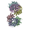
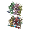
| Title | Crystal structure of nitrite and NO bound three-domain copper-containing nitrite reductase from Hyphomicrobium denitrificans strain 1NES1 | ||||||
 Components Components | Copper-containing nitrite reductase | ||||||
 Keywords Keywords | OXIDOREDUCTASE / Copper-containing nitrite reductase / Hyphomicrobium denitrificans strain 1NES1 / electron transfer | ||||||
| Function / homology |  Function and homology information Function and homology informationnitrite reductase (NO-forming) / nitrite reductase (NO-forming) activity / electron transfer activity / copper ion binding Similarity search - Function | ||||||
| Biological species |  Hyphomicrobium denitrificans 1NES1 (bacteria) Hyphomicrobium denitrificans 1NES1 (bacteria) | ||||||
| Method |  X-RAY DIFFRACTION / SYNCHROTRON / MOLECULAR REPLACEMENT / Resolution: 2.25 Å X-RAY DIFFRACTION / SYNCHROTRON / MOLECULAR REPLACEMENT / Resolution: 2.25 Å | ||||||
 Authors Authors | Sasaki, D. / Watanabe, T.F. / Eady, R.R. / Garratt, R.C. / Antonyuk, S.V. / Hasnain, S.S. | ||||||
| Funding support |  United Kingdom, 1items United Kingdom, 1items
| ||||||
 Citation Citation | Journal: Iucrj / Year: 2020 Title: Structures of substrate- and product-bound forms of a multi-domain copper nitrite reductase shed light on the role of domain tethering in protein complexes. Authors: Sasaki, D. / Watanabe, T.F. / Eady, R.R. / Garratt, R.C. / Antonyuk, S.V. / Hasnain, S.S. | ||||||
| History |
|
- Structure visualization
| Structure viewer | Molecule: MolmilJmol/JSmol |
|---|
- Downloads & links
-Download
| PDBx/mmCIF format | 6tfd.cif.gz | 265 KB | Display | PDBx/mmCIF format |
|---|---|---|---|---|
| PDB format | pdb6tfd.ent.gz | 211.9 KB | Display | PDB format |
| PDBx/mmJSON format | 6tfd.json.gz | Tree view | PDBx/mmJSON format | |
| Others |  Other downloads Other downloads |
-Validation report
| Arichive directory | https://data.pdbj.org/pub/pdb/validation_reports/tf/6tfdftp://data.pdbj.org/pub/pdb/validation_reports/tf/6tfd | HTTPS FTP |
|---|
-Related structure data
| Related structure data | 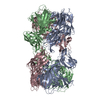 6tfoC 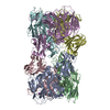 2dv6S S: Starting model for refinement C: citing same article ( |
|---|---|
| Similar structure data |


-Links
 PDBj
PDBj


- Assembly
| Deposited unit | 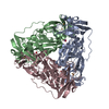
| ||||||||
|---|---|---|---|---|---|---|---|---|---|
| 1 | 
| ||||||||
| Unit cell |
|
-Components
| #1: Protein | Mass: 49050.926 Da / Num. of mol.: 3 Source method: isolated from a genetically manipulated source Source: (gene. exp.) Hyphomicrobium denitrificans 1NES1 (bacteria)Gene: HYPDE_25578 / Plasmid: pET-26b(+) / Production host: #2: Chemical | ChemComp-CU /   Mass: 63.546 Da / Num. of mol.: 9 / Source method: obtained synthetically / Formula: Cu Mass: 63.546 Da / Num. of mol.: 9 / Source method: obtained synthetically / Formula: Cu#3: Chemical | 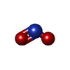  Mass: 46.005 Da / Num. of mol.: 2 / Source method: obtained synthetically / Formula: NO2 / Feature type: SUBJECT OF INVESTIGATION Mass: 46.005 Da / Num. of mol.: 2 / Source method: obtained synthetically / Formula: NO2 / Feature type: SUBJECT OF INVESTIGATION#4: Chemical | ChemComp-NO / |   Mass: 30.006 Da / Num. of mol.: 1 / Source method: obtained synthetically / Formula: NO / Feature type: SUBJECT OF INVESTIGATION Mass: 30.006 Da / Num. of mol.: 1 / Source method: obtained synthetically / Formula: NO / Feature type: SUBJECT OF INVESTIGATION#5: Water | ChemComp-HOH / |  Mass: 18.015 Da / Num. of mol.: 639 / Source method: isolated from a natural source / Formula: H2O Mass: 18.015 Da / Num. of mol.: 639 / Source method: isolated from a natural source / Formula: H2OHas ligand of interest | Y | |
|---|
-Experimental details
-Experiment
| Experiment | Method: X-RAY DIFFRACTION / Number of used crystals: 1 |
|---|
- Sample preparation
| Crystal | Density Matthews: 2.25 Å3/Da / Density % sol: 45.35 % / Description: Hexagonal |
|---|---|
| Crystal grow | Temperature: 298 K / Method: vapor diffusion, hanging drop / pH: 5.3 Details: 22.5% (v/v) PEG Smear Low, 0.1 M Sodium cacodylate pH5.3 0.2 M Ammonium nitrate |
-Data collection
| Diffraction | Mean temperature: 100 K / Serial crystal experiment: N |
|---|---|
| Diffraction source | Source: SYNCHROTRON / Site: Diamond / Beamline: I04 / Wavelength: 0.9795 Å |
| Detector | Type: DECTRIS EIGER X 16M / Detector: PIXEL / Date: Jun 29, 2019 |
| Radiation | Protocol: SINGLE WAVELENGTH / Monochromatic (M) / Laue (L): M / Scattering type: x-ray |
| Radiation wavelength | Wavelength: 0.9795 Å / Relative weight: 1 |
| Reflection | Resolution: 2.1→126.37 Å / Num. obs: 81889 / % possible obs: 100 % / Redundancy: 12.1 % / Biso Wilson estimate: 45.07 Å2 / CC1/2: 0.998 / Rmerge(I) obs: 0.216 / Rpim(I) all: 0.064 / Net I/σ(I): 7.7 |
| Reflection shell | Resolution: 2.1→2.14 Å / Redundancy: 12.3 % / Rmerge(I) obs: 2.845 / Mean I/σ(I) obs: 0.855 / Num. unique obs: 3922 / CC1/2: 0.4256 / Rpim(I) all: 0.828 / % possible all: 100 |
- Processing
| Software |
| ||||||||||||||||||||||||||||||||||||||||||||||||||||||||||||
|---|---|---|---|---|---|---|---|---|---|---|---|---|---|---|---|---|---|---|---|---|---|---|---|---|---|---|---|---|---|---|---|---|---|---|---|---|---|---|---|---|---|---|---|---|---|---|---|---|---|---|---|---|---|---|---|---|---|---|---|---|---|
| Refinement | Method to determine structure: MOLECULAR REPLACEMENT Starting model: 2DV6 Resolution: 2.25→126.37 Å / Cor.coef. Fo:Fc: 0.971 / Cor.coef. Fo:Fc free: 0.951 / SU B: 9.093 / SU ML: 0.199 / Cross valid method: THROUGHOUT / σ(F): 0 / ESU R: 0.266 / ESU R Free: 0.209 Details: HYDROGENS HAVE BEEN ADDED IN THE RIDING POSITIONS U VALUES : REFINED INDIVIDUALLY
| ||||||||||||||||||||||||||||||||||||||||||||||||||||||||||||
| Solvent computation | Ion probe radii: 0.8 Å / Shrinkage radii: 0.8 Å / VDW probe radii: 1.2 Å | ||||||||||||||||||||||||||||||||||||||||||||||||||||||||||||
| Displacement parameters | Biso max: 125.29 Å2 / Biso mean: 50.816 Å2 / Biso min: 30.43 Å2
| ||||||||||||||||||||||||||||||||||||||||||||||||||||||||||||
| Refinement step | Cycle: final / Resolution: 2.25→126.37 Å
| ||||||||||||||||||||||||||||||||||||||||||||||||||||||||||||
| Refine LS restraints |
| ||||||||||||||||||||||||||||||||||||||||||||||||||||||||||||
| LS refinement shell | Resolution: 2.25→2.308 Å / Rfactor Rfree error: 0
|
