 Movie
Movie Controller

[English] 日本語
 Yorodumi
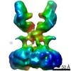
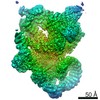
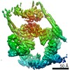
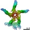
- PDB-6tda: Structure of SWI/SNF chromatin remodeler RSC bound to a nucleosome -
+ Open data

- Basic information
| Entry | Database: PDB / ID: 6tda | ||||||||||||
|---|---|---|---|---|---|---|---|---|---|---|---|---|---|
| Title | Structure of SWI/SNF chromatin remodeler RSC bound to a nucleosome | ||||||||||||
 Components Components |
| ||||||||||||
 Keywords Keywords | DNA BINDING PROTEIN / Chromatin remodeler DNA binding Nucleosome binding ATPase Transcription | ||||||||||||
| Function / homology |  Function and homology information Function and homology informationhistone H2A reader activity / RHO GTPases activate IQGAPs / RHO GTPases Activate WASPs and WAVEs / : / : / : / Regulation of actin dynamics for phagocytic cup formation / regulation of nuclear cell cycle DNA replication / histone H3 reader activity / Platelet degranulation ...histone H2A reader activity / RHO GTPases activate IQGAPs / RHO GTPases Activate WASPs and WAVEs / : / : / : / Regulation of actin dynamics for phagocytic cup formation / regulation of nuclear cell cycle DNA replication / histone H3 reader activity / Platelet degranulation / CENP-A containing chromatin assembly / DNA translocase activity / histone H3K14ac reader activity / nucleosome array spacer activity / RSC-type complex / nucleosome disassembly / ATP-dependent chromatin remodeler activity / SWI/SNF complex / nuclear chromosome / NuA4 histone acetyltransferase complex / rRNA transcription / histone H4 reader activity / chromosome, centromeric region / nucleosome binding / cytoskeleton organization / DNA helicase activity / helicase activity / transcription coregulator activity / positive regulation of transcription elongation by RNA polymerase II / chromosome segregation / transcription elongation by RNA polymerase II / meiotic cell cycle / chromatin DNA binding / base-excision repair / double-strand break repair via nonhomologous end joining / G2/M transition of mitotic cell cycle / structural constituent of chromatin / nucleosome / double-strand break repair / nucleosome assembly / heterochromatin formation / chromatin organization / histone binding / DNA helicase / chromatin remodeling / protein heterodimerization activity / chromatin binding / regulation of transcription by RNA polymerase II / regulation of DNA-templated transcription / positive regulation of DNA-templated transcription / chromatin / structural molecule activity / positive regulation of transcription by RNA polymerase II / ATP hydrolysis activity / DNA binding / zinc ion binding / nucleoplasm / ATP binding / nucleus Similarity search - Function | ||||||||||||
| Biological species |  synthetic construct (others) | ||||||||||||
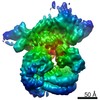
| Method | ELECTRON MICROSCOPY / single particle reconstruction / cryo EM / Resolution: 15 Å | ||||||||||||
 Authors Authors | Wagner, F.R. / Dienemann, C. / Wang, H. / Stuetzer, A. / Tegunov, D. / Urlaub, H. / Cramer, P. | ||||||||||||
| Funding support | 3items
| ||||||||||||
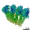
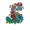
 Citation Citation | Journal: Nature / Year: 2020 Title: Structure of SWI/SNF chromatin remodeller RSC bound to a nucleosome. Authors: Felix R Wagner / Christian Dienemann / Haibo Wang / Alexandra Stützer / Dimitry Tegunov / Henning Urlaub / Patrick Cramer /  Abstract: Chromatin-remodelling complexes of the SWI/SNF family function in the formation of nucleosome-depleted, transcriptionally active promoter regions (NDRs). In the yeast Saccharomyces cerevisiae, the ...Chromatin-remodelling complexes of the SWI/SNF family function in the formation of nucleosome-depleted, transcriptionally active promoter regions (NDRs). In the yeast Saccharomyces cerevisiae, the essential SWI/SNF complex RSC contains 16 subunits, including the ATP-dependent DNA translocase Sth1. RSC removes nucleosomes from promoter regions and positions the specialized +1 and -1 nucleosomes that flank NDRs. Here we present the cryo-electron microscopy structure of RSC in complex with a nucleosome substrate. The structure reveals that RSC forms five protein modules and suggests key features of the remodelling mechanism. The body module serves as a scaffold for the four flexible modules that we call DNA-interacting, ATPase, arm and actin-related protein (ARP) modules. The DNA-interacting module binds extra-nucleosomal DNA and is involved in the recognition of promoter DNA elements that influence RSC functionality. The ATPase and arm modules sandwich the nucleosome disc with the Snf2 ATP-coupling (SnAC) domain and the finger helix, respectively. The translocase motor of the ATPase module engages with the edge of the nucleosome at superhelical location +2. The mobile ARP module may modulate translocase-nucleosome interactions to regulate RSC activity. The RSC-nucleosome structure provides a basis for understanding NDR formation and the structure and function of human SWI/SNF complexes that are frequently mutated in cancer. | ||||||||||||
| History |
|
- Structure visualization
| Movie |
Movie viewer |
|---|---|
| Structure viewer | Molecule: MolmilJmol/JSmol |
- Downloads & links
-Download
| PDBx/mmCIF format | 6tda.cif.gz | 1 MB | Display | PDBx/mmCIF format |
|---|---|---|---|---|
| PDB format | pdb6tda.ent.gz | 798 KB | Display | PDB format |
| PDBx/mmJSON format | 6tda.json.gz | Tree view | PDBx/mmJSON format | |
| Others |  Other downloads Other downloads |
-Validation report
| Arichive directory | https://data.pdbj.org/pub/pdb/validation_reports/td/6tdaftp://data.pdbj.org/pub/pdb/validation_reports/td/6tda | HTTPS FTP |
|---|
-Related structure data
| Related structure data |  10465MC M: map data used to model this data C: citing same article ( |
|---|---|
| Similar structure data |
-Links
 PDBj
PDBj


- Assembly
| Deposited unit | 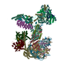
|
|---|---|
| 1 |
|
-Components
-Protein , 10 types, 14 molecules AEBFCGDHQSTUVX
| #1: Protein | Mass: 15303.930 Da / Num. of mol.: 2 Source method: isolated from a genetically manipulated source Source: (gene. exp.)  #2: Protein | Mass: 11263.231 Da / Num. of mol.: 2 Source method: isolated from a genetically manipulated source Source: (gene. exp.) #3: Protein | Mass: 13978.241 Da / Num. of mol.: 2 Source method: isolated from a genetically manipulated source Source: (gene. exp.) #4: Protein | Mass: 13524.752 Da / Num. of mol.: 2 Source method: isolated from a genetically manipulated source Source: (gene. exp.) #13: Protein | | Mass: 9192.524 Da / Num. of mol.: 1 / Source method: isolated from a natural source Source: (natural) References: UniProt: Q9URQ5 #15: Protein | | Mass: 156982.406 Da / Num. of mol.: 1 / Source method: isolated from a natural source Source: (natural) References: UniProt: P32597, DNA helicase #16: Protein | | Mass: 54613.332 Da / Num. of mol.: 1 / Source method: isolated from a natural source Source: (natural) References: UniProt: Q12406 #17: Protein | | Mass: 53366.398 Da / Num. of mol.: 1 / Source method: isolated from a natural source Source: (natural) References: UniProt: Q05123 #18: Protein | | Mass: 18005.195 Da / Num. of mol.: 1 / Source method: isolated from a natural source Source: (natural) References: UniProt: P53330 #19: Protein | | Mass: 32613.104 Da / Num. of mol.: 1 / Source method: isolated from a natural source / Source: (natural) |
|---|
-DNA chain , 2 types, 2 molecules IJ
| #5: DNA chain | Mass: 72876.344 Da / Num. of mol.: 1 / Source method: obtained synthetically / Source: (synth.) synthetic construct (others) |
|---|---|
| #6: DNA chain | Mass: 73478.852 Da / Num. of mol.: 1 / Source method: obtained synthetically / Source: (synth.) synthetic construct (others) |
-Chromatin structure-remodeling complex subunit ... , 4 types, 4 molecules KMNR
| #7: Protein | Mass: 48833.180 Da / Num. of mol.: 1 / Source method: isolated from a natural source Source: (natural) References: UniProt: Q06168 |
|---|---|
| #9: Protein | Mass: 49716.520 Da / Num. of mol.: 1 / Source method: isolated from a natural source Source: (natural) References: UniProt: P32832 |
| #10: Protein | Mass: 65289.309 Da / Num. of mol.: 1 / Source method: isolated from a natural source Source: (natural) References: UniProt: Q03124 |
| #14: Protein | Mass: 72372.375 Da / Num. of mol.: 1 / Source method: isolated from a natural source Source: (natural) References: UniProt: Q02206 |
-Chromatin structure-remodeling complex protein ... , 3 types, 3 molecules LOP
| #8: Protein | Mass: 126489.883 Da / Num. of mol.: 1 / Source method: isolated from a natural source Source: (natural) References: UniProt: P43609 |
|---|---|
| #11: Protein | Mass: 54222.691 Da / Num. of mol.: 1 / Source method: isolated from a natural source Source: (natural) References: UniProt: P25632 |
| #12: Protein | Mass: 57871.309 Da / Num. of mol.: 1 / Source method: isolated from a natural source Source: (natural) References: UniProt: Q07979 |
-Non-polymers , 1 types, 1 molecules 
| #20: Chemical | ChemComp-ZN /  Mass: 65.409 Da / Num. of mol.: 1 / Source method: obtained synthetically / Formula: Zn Mass: 65.409 Da / Num. of mol.: 1 / Source method: obtained synthetically / Formula: Zn |
|---|
-Details
| Has ligand of interest | N |
|---|---|
| Has protein modification | Y |
-Experimental details
-Experiment
| Experiment | Method: ELECTRON MICROSCOPY |
|---|---|
| EM experiment | Aggregation state: PARTICLE / 3D reconstruction method: single particle reconstruction |
- Sample preparation
| Component |
| |||||||||||||||||||||||||||||||||||
|---|---|---|---|---|---|---|---|---|---|---|---|---|---|---|---|---|---|---|---|---|---|---|---|---|---|---|---|---|---|---|---|---|---|---|---|---|
| Source (natural) |
| |||||||||||||||||||||||||||||||||||
| Source (recombinant) |
| |||||||||||||||||||||||||||||||||||
| Buffer solution | pH: 7.6 | |||||||||||||||||||||||||||||||||||
| Specimen | Embedding applied: NO / Shadowing applied: NO / Staining applied: NO / Vitrification applied: YES | |||||||||||||||||||||||||||||||||||
| Vitrification | Cryogen name: ETHANE |
- Electron microscopy imaging
| Experimental equipment |  Model: Titan Krios / Image courtesy: FEI Company |
|---|---|
| Microscopy | Model: FEI TITAN KRIOS |
| Electron gun | Electron source:  FIELD EMISSION GUN / Accelerating voltage: 300 kV / Illumination mode: FLOOD BEAM FIELD EMISSION GUN / Accelerating voltage: 300 kV / Illumination mode: FLOOD BEAM |
| Electron lens | Mode: BRIGHT FIELD |
| Image recording | Electron dose: 45.4 e/Å2 / Film or detector model: GATAN K2 SUMMIT (4k x 4k) |
- Processing
| CTF correction | Type: PHASE FLIPPING AND AMPLITUDE CORRECTION |
|---|---|
| 3D reconstruction | Resolution: 15 Å / Resolution method: OTHER / Num. of particles: 372442 / Symmetry type: POINT |
