 Movie
Movie Controller

[English] 日本語
 Yorodumi
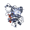
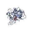
- PDB-6tbq: AA13 Lytic polysaccharide monooxygenase from Aspergillus oryzae p... -
+ Open data

- Basic information
| Entry | Database: PDB / ID: 6tbq | ||||||||||||
|---|---|---|---|---|---|---|---|---|---|---|---|---|---|
| Title | AA13 Lytic polysaccharide monooxygenase from Aspergillus oryzae partially in Cu(II) state | ||||||||||||
 Components Components | AoAA13 | ||||||||||||
 Keywords Keywords | OXIDOREDUCTASE / beta-sandwich fold / low dose | ||||||||||||
| Function / homology | : / Cellulose/chitin-binding protein, N-terminal / Lytic polysaccharide mono-oxygenase, cellulose-degrading / membrane / metal ion binding / COPPER (II) ION / Inactive AA13 family lytic polysaccharide monooxygenase Function and homology information Function and homology information | ||||||||||||
| Biological species |  | ||||||||||||
| Method |  X-RAY DIFFRACTION / SYNCHROTRON / FOURIER SYNTHESIS / Resolution: 1.72 Å X-RAY DIFFRACTION / SYNCHROTRON / FOURIER SYNTHESIS / Resolution: 1.72 Å | ||||||||||||
 Authors Authors | Muderspach, S.J. / Lo Leggio, L. / Tandrup, T. / Frandsen, K.E.H. / Poulsen, J.C.N. | ||||||||||||
| Funding support |  Denmark, 3items Denmark, 3items
| ||||||||||||
 Citation Citation | Journal: Amylase / Year: 2019 Title: Further structural studies of the lytic polysaccharide monooxygenase AoAA13 belonging to the starch-active AA13 family Authors: Muderspach, S.J. / Tandrup, T. / Frandsen, K.E.H. / Santoni, G. / Poulsen, J.C.N. / Lo Leggio, L. | ||||||||||||
| History |
|
- Structure visualization
| Structure viewer | Molecule: MolmilJmol/JSmol |
|---|
- Downloads & links
-Download
| PDBx/mmCIF format | 6tbq.cif.gz | 64.7 KB | Display | PDBx/mmCIF format |
|---|---|---|---|---|
| PDB format | pdb6tbq.ent.gz | 45.4 KB | Display | PDB format |
| PDBx/mmJSON format | 6tbq.json.gz | Tree view | PDBx/mmJSON format | |
| Others |  Other downloads Other downloads |
-Validation report
| Arichive directory | https://data.pdbj.org/pub/pdb/validation_reports/tb/6tbqftp://data.pdbj.org/pub/pdb/validation_reports/tb/6tbq | HTTPS FTP |
|---|
-Related structure data
| Related structure data |  6tbrC  6tc4C  4opbS S: Starting model for refinement C: citing same article ( |
|---|---|
| Similar structure data |


-Links
 PDBj
PDBj- Assembly
| Deposited unit | 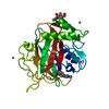
| ||||||||
|---|---|---|---|---|---|---|---|---|---|
| 1 |
| ||||||||
| Unit cell |
|
-Components
| #1: Protein | Mass: 25691.057 Da / Num. of mol.: 1 Source method: isolated from a genetically manipulated source Source: (gene. exp.) | ||||
|---|---|---|---|---|---|
| #2: Chemical | ChemComp-CU /   Mass: 63.546 Da / Num. of mol.: 1 Mass: 63.546 Da / Num. of mol.: 1Source method: isolated from a genetically manipulated source Formula: Cu / Feature type: SUBJECT OF INVESTIGATION | ||||
| #3: Sugar | ChemComp-NAG /   Type: D-saccharide, beta linking / Mass: 221.208 Da / Num. of mol.: 1 Type: D-saccharide, beta linking / Mass: 221.208 Da / Num. of mol.: 1Source method: isolated from a genetically manipulated source Formula: C8H15NO6 | ||||
| #4: Chemical | ChemComp-ZN /   Mass: 65.409 Da / Num. of mol.: 5 / Source method: obtained synthetically / Formula: Zn Mass: 65.409 Da / Num. of mol.: 5 / Source method: obtained synthetically / Formula: Zn#5: Water | ChemComp-HOH / |  Mass: 18.015 Da / Num. of mol.: 160 / Source method: isolated from a natural source / Formula: H2O Mass: 18.015 Da / Num. of mol.: 160 / Source method: isolated from a natural source / Formula: H2OHas ligand of interest | Y | |
-Experimental details
-Experiment
| Experiment | Method: X-RAY DIFFRACTION / Number of used crystals: 1 |
|---|
- Sample preparation
| Crystal | Density Matthews: 2.02 Å3/Da / Density % sol: 39.03 % |
|---|---|
| Crystal grow | Temperature: 298 K / Method: vapor diffusion, hanging drop / pH: 5 Details: 0.1 M buffer system (II) (0.2 M L-malic acid, 0.04 M MES, 0.4 M Tris) pH 5, 0.2 M Zn-acetate, 23% PEG 3000. The crystals were soaked in 0.09 M Buffer system (II) pH 5, 22.5% PEG 3000, 0.5 M ...Details: 0.1 M buffer system (II) (0.2 M L-malic acid, 0.04 M MES, 0.4 M Tris) pH 5, 0.2 M Zn-acetate, 23% PEG 3000. The crystals were soaked in 0.09 M Buffer system (II) pH 5, 22.5% PEG 3000, 0.5 M Cu-acetate, 1 mg/ml BSA Temp details: Room temperature |
-Data collection
| Diffraction | Mean temperature: 100 K / Serial crystal experiment: N |
|---|---|
| Diffraction source | Source: SYNCHROTRON / Site: ESRF  / Beamline: ID23-1 / Wavelength: 1.3 Å / Beamline: ID23-1 / Wavelength: 1.3 Å |
| Detector | Type: DECTRIS PILATUS 6M-F / Detector: PIXEL / Date: Jun 25, 2017 |
| Radiation | Protocol: SINGLE WAVELENGTH / Monochromatic (M) / Laue (L): M / Scattering type: x-ray |
| Radiation wavelength | Wavelength: 1.3 Å / Relative weight: 1 |
| Reflection | Resolution: 1.72→46.9 Å / Num. obs: 21830 / % possible obs: 99.1 % / Redundancy: 3.77 % / CC1/2: 0.987 / Rrim(I) all: 0.332 / Net I/σ(I): 4.6 |
| Reflection shell | Resolution: 1.72→1.76 Å / Mean I/σ(I) obs: 0.6 / Num. unique obs: 758 / CC1/2: 0.425 / Rrim(I) all: 2.327 |
- Processing
| Software |
| ||||||||||||||||||||||||||||||||||||||||||||||||||||||||||||
|---|---|---|---|---|---|---|---|---|---|---|---|---|---|---|---|---|---|---|---|---|---|---|---|---|---|---|---|---|---|---|---|---|---|---|---|---|---|---|---|---|---|---|---|---|---|---|---|---|---|---|---|---|---|---|---|---|---|---|---|---|---|
| Refinement | Method to determine structure: FOURIER SYNTHESIS Starting model: 4OPB Resolution: 1.72→46.9 Å / Cor.coef. Fo:Fc: 0.952 / Cor.coef. Fo:Fc free: 0.935 / SU B: 6.026 / SU ML: 0.174 / Cross valid method: THROUGHOUT / σ(F): 0 / ESU R: 0.146 / ESU R Free: 0.139
| ||||||||||||||||||||||||||||||||||||||||||||||||||||||||||||
| Solvent computation | Ion probe radii: 0.8 Å / Shrinkage radii: 0.8 Å / VDW probe radii: 1.2 Å | ||||||||||||||||||||||||||||||||||||||||||||||||||||||||||||
| Displacement parameters | Biso max: 68.98 Å2 / Biso mean: 24.441 Å2 / Biso min: 13.57 Å2
| ||||||||||||||||||||||||||||||||||||||||||||||||||||||||||||
| Refinement step | Cycle: final / Resolution: 1.72→46.9 Å
| ||||||||||||||||||||||||||||||||||||||||||||||||||||||||||||
| Refine LS restraints |
| ||||||||||||||||||||||||||||||||||||||||||||||||||||||||||||
| LS refinement shell | Resolution: 1.72→1.762 Å / Rfactor Rfree error: 0
|
