 Movie
Movie Controller

[English] 日本語
 Yorodumi
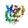
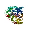
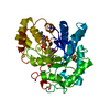
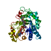
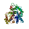
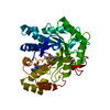
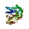
- PDB-6t7q: Human Aldose Reductase Mutant L301A in Complex with a Ligand with... -
+ Open data

- Basic information
| Entry | Database: PDB / ID: 6t7q | ||||||
|---|---|---|---|---|---|---|---|
| Title | Human Aldose Reductase Mutant L301A in Complex with a Ligand with an IDD Structure (3-({[2-(carboxymethoxy)-4-fluorobenzoyl]amino}methyl)benzoic acid) | ||||||
 Components Components | Aldo-keto reductase family 1 member B1 | ||||||
 Keywords Keywords | OXIDOREDUCTASE / L301A Mutant / SAR_061 ligand / transient binding pocket | ||||||
| Function / homology |  Function and homology information Function and homology informationglyceraldehyde oxidoreductase activity / Fructose biosynthesis / fructose biosynthetic process / L-glucuronate reductase activity / aldose reductase / D/L-glyceraldehyde reductase / glycerol dehydrogenase (NADP+) activity / C21-steroid hormone biosynthetic process / NADP-retinol dehydrogenase / Pregnenolone biosynthesis ...glyceraldehyde oxidoreductase activity / Fructose biosynthesis / fructose biosynthetic process / L-glucuronate reductase activity / aldose reductase / D/L-glyceraldehyde reductase / glycerol dehydrogenase (NADP+) activity / C21-steroid hormone biosynthetic process / NADP-retinol dehydrogenase / Pregnenolone biosynthesis / allyl-alcohol dehydrogenase / allyl-alcohol dehydrogenase activity / Galactose catabolism / prostaglandin H2 endoperoxidase reductase activity / regulation of urine volume / all-trans-retinol dehydrogenase (NADP+) activity / metanephric collecting duct development / daunorubicin metabolic process / doxorubicin metabolic process / retinal dehydrogenase (NAD+) activity / epithelial cell maturation / aldose reductase (NADPH) activity / cellular hyperosmotic salinity response / renal water homeostasis / retinoid metabolic process / carbohydrate metabolic process / electron transfer activity / negative regulation of apoptotic process / mitochondrion / : / extracellular exosome / nucleoplasm / cytosol Similarity search - Function | ||||||
| Biological species |  Homo sapiens (human) Homo sapiens (human) | ||||||
| Method |  X-RAY DIFFRACTION / SYNCHROTRON / MOLECULAR REPLACEMENT / Resolution: 1.01 Å X-RAY DIFFRACTION / SYNCHROTRON / MOLECULAR REPLACEMENT / Resolution: 1.01 Å | ||||||
 Authors Authors | Hubert, L.-S. / Ley, M. / Heine, A. / Klebe, G. | ||||||
 Citation Citation | Journal: To Be Published Title: Human Aldose Reductase Mutant L301A in Complex with a Ligand with an IDD Structure (3-({[2-(carboxymethoxy)-4-fluorobenzoyl]amino}methyl)benzoic acid) Authors: Hubert, L.-S. / Ley, M. / Scheer, F. / Diederich, W. / Heine, A. / Klebe, G. | ||||||
| History |
|
- Structure visualization
| Structure viewer | Molecule: MolmilJmol/JSmol |
|---|
- Downloads & links
-Download
| PDBx/mmCIF format | 6t7q.cif.gz | 271.9 KB | Display | PDBx/mmCIF format |
|---|---|---|---|---|
| PDB format | pdb6t7q.ent.gz | 185.5 KB | Display | PDB format |
| PDBx/mmJSON format | 6t7q.json.gz | Tree view | PDBx/mmJSON format | |
| Others |  Other downloads Other downloads |
-Validation report
| Arichive directory | https://data.pdbj.org/pub/pdb/validation_reports/t7/6t7qftp://data.pdbj.org/pub/pdb/validation_reports/t7/6t7q | HTTPS FTP |
|---|
-Related structure data
| Related structure data | 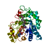 4prrS S: Starting model for refinement |
|---|---|
| Similar structure data |


-Links
 PDBj
PDBj
- Assembly
| Deposited unit | 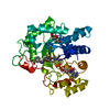
| ||||||||||||
|---|---|---|---|---|---|---|---|---|---|---|---|---|---|
| 1 |
| ||||||||||||
| Unit cell |
|
-Components
| #1: Protein | Mass: 35856.262 Da / Num. of mol.: 1 / Mutation: L301A Source method: isolated from a genetically manipulated source Source: (gene. exp.) Homo sapiens (human) / Gene: AKR1B1, ALDR1, ALR2 / Plasmid: pET15b / Production host:  References: UniProt: P15121, NADP-retinol dehydrogenase, D/L-glyceraldehyde reductase, allyl-alcohol dehydrogenase, aldose reductase |
|---|---|
| #2: Chemical | ChemComp-4G7 / 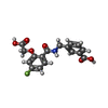  Mass: 347.295 Da / Num. of mol.: 1 / Source method: obtained synthetically / Formula: C17H14FNO6 / Feature type: SUBJECT OF INVESTIGATION Mass: 347.295 Da / Num. of mol.: 1 / Source method: obtained synthetically / Formula: C17H14FNO6 / Feature type: SUBJECT OF INVESTIGATION |
| #3: Chemical | ChemComp-CIT /   Mass: 192.124 Da / Num. of mol.: 1 / Source method: obtained synthetically / Formula: C6H8O7 Mass: 192.124 Da / Num. of mol.: 1 / Source method: obtained synthetically / Formula: C6H8O7 |
| #4: Chemical | ChemComp-NAP /   Mass: 743.405 Da / Num. of mol.: 1 / Source method: obtained synthetically / Formula: C21H28N7O17P3 Mass: 743.405 Da / Num. of mol.: 1 / Source method: obtained synthetically / Formula: C21H28N7O17P3 |
| #5: Water | ChemComp-HOH /  Mass: 18.015 Da / Num. of mol.: 384 / Source method: isolated from a natural source / Formula: H2O Mass: 18.015 Da / Num. of mol.: 384 / Source method: isolated from a natural source / Formula: H2O |
| Has ligand of interest | Y |
-Experimental details
-Experiment
| Experiment | Method: X-RAY DIFFRACTION / Number of used crystals: 1 |
|---|
- Sample preparation
| Crystal | Density Matthews: 2.18 Å3/Da / Density % sol: 43.67 % |
|---|---|
| Crystal grow | Temperature: 291 K / Method: vapor diffusion, hanging drop / pH: 5 Details: 50 mM Di-Ammoniumhydrogen citrate pH 5.0: 15 mg/mL hAR, 5.2 mg/mL DTT, 0.7 mg/mL NADP+, 5% (w/v) PEG 6000 Reservoir: 120 mM Di-Ammoniumhydrogen citrate pH 5.0, 20% (w/v) PEG 6000 Soaking- ...Details: 50 mM Di-Ammoniumhydrogen citrate pH 5.0: 15 mg/mL hAR, 5.2 mg/mL DTT, 0.7 mg/mL NADP+, 5% (w/v) PEG 6000 Reservoir: 120 mM Di-Ammoniumhydrogen citrate pH 5.0, 20% (w/v) PEG 6000 Soaking-buffer: 120 mM Di-Ammoniumhydrogen citrate pH 5.0, 25% (w/v) PEG 6000 |
-Data collection
| Diffraction | Mean temperature: 100 K / Serial crystal experiment: N |
|---|---|
| Diffraction source | Source: SYNCHROTRON / Site: BESSY  / Beamline: 14.1 / Wavelength: 0.9184 Å / Beamline: 14.1 / Wavelength: 0.9184 Å |
| Detector | Type: DECTRIS PILATUS 6M / Detector: PIXEL / Date: Jan 19, 2018 |
| Radiation | Protocol: SINGLE WAVELENGTH / Monochromatic (M) / Laue (L): M / Scattering type: x-ray |
| Radiation wavelength | Wavelength: 0.9184 Å / Relative weight: 1 |
| Reflection | Resolution: 1.007→39.84 Å / Num. obs: 155904 / % possible obs: 95.3 % / Redundancy: 3.37 % / Biso Wilson estimate: 6.55 Å2 / CC1/2: 0.998 / Rsym value: 0.052 / Net I/σ(I): 13.84 |
| Reflection shell | Resolution: 1.01→1.07 Å / Redundancy: 3.17 % / Mean I/σ(I) obs: 2.47 / Num. unique obs: 23595 / CC1/2: 0.876 / Rsym value: 0.481 / % possible all: 89.4 |
- Processing
| Software |
| |||||||||||||||||||||||||||||||||||||||||||||||||||||||||||||||||||||||||||||||||||||||||||||||||||||||||||||||||||||||||||||||||||||||||||||||||||||||||||||||||||||||||||||||||||||||||||||||||||||||||||||||||||||||||
|---|---|---|---|---|---|---|---|---|---|---|---|---|---|---|---|---|---|---|---|---|---|---|---|---|---|---|---|---|---|---|---|---|---|---|---|---|---|---|---|---|---|---|---|---|---|---|---|---|---|---|---|---|---|---|---|---|---|---|---|---|---|---|---|---|---|---|---|---|---|---|---|---|---|---|---|---|---|---|---|---|---|---|---|---|---|---|---|---|---|---|---|---|---|---|---|---|---|---|---|---|---|---|---|---|---|---|---|---|---|---|---|---|---|---|---|---|---|---|---|---|---|---|---|---|---|---|---|---|---|---|---|---|---|---|---|---|---|---|---|---|---|---|---|---|---|---|---|---|---|---|---|---|---|---|---|---|---|---|---|---|---|---|---|---|---|---|---|---|---|---|---|---|---|---|---|---|---|---|---|---|---|---|---|---|---|---|---|---|---|---|---|---|---|---|---|---|---|---|---|---|---|---|---|---|---|---|---|---|---|---|---|---|---|---|---|---|---|---|
| Refinement | Method to determine structure: MOLECULAR REPLACEMENT Starting model: 4PRR Resolution: 1.01→39.84 Å / SU ML: 0.1092 / Cross valid method: FREE R-VALUE / σ(F): 1.33 / Phase error: 12.0585 Stereochemistry target values: GeoStd + Monomer Library + CDL v1.2
| |||||||||||||||||||||||||||||||||||||||||||||||||||||||||||||||||||||||||||||||||||||||||||||||||||||||||||||||||||||||||||||||||||||||||||||||||||||||||||||||||||||||||||||||||||||||||||||||||||||||||||||||||||||||||
| Solvent computation | Shrinkage radii: 0.9 Å / VDW probe radii: 1.11 Å / Solvent model: FLAT BULK SOLVENT MODEL | |||||||||||||||||||||||||||||||||||||||||||||||||||||||||||||||||||||||||||||||||||||||||||||||||||||||||||||||||||||||||||||||||||||||||||||||||||||||||||||||||||||||||||||||||||||||||||||||||||||||||||||||||||||||||
| Displacement parameters | Biso mean: 11.81 Å2 | |||||||||||||||||||||||||||||||||||||||||||||||||||||||||||||||||||||||||||||||||||||||||||||||||||||||||||||||||||||||||||||||||||||||||||||||||||||||||||||||||||||||||||||||||||||||||||||||||||||||||||||||||||||||||
| Refinement step | Cycle: LAST / Resolution: 1.01→39.84 Å
| |||||||||||||||||||||||||||||||||||||||||||||||||||||||||||||||||||||||||||||||||||||||||||||||||||||||||||||||||||||||||||||||||||||||||||||||||||||||||||||||||||||||||||||||||||||||||||||||||||||||||||||||||||||||||
| Refine LS restraints |
| |||||||||||||||||||||||||||||||||||||||||||||||||||||||||||||||||||||||||||||||||||||||||||||||||||||||||||||||||||||||||||||||||||||||||||||||||||||||||||||||||||||||||||||||||||||||||||||||||||||||||||||||||||||||||
| LS refinement shell |
|
