- PDB-6t6r: Human endoplasmic reticulum aminopeptidase 1 (ERAP1) in complex w... -
+
Open data
ID or keywords:
Loading...
-
Basic information
Entry
Database: PDB / ID: 6t6r
Title
Human endoplasmic reticulum aminopeptidase 1 (ERAP1) in complex with (4aR,5S,6R,8S,8aR)-5-(2-(Furan-3-yl)ethyl)-8-hydroxy-5,6,8a-trimethyl-3,4,4a,5,6,7,8,8a-octahydronaphthalene-1-carboxylic acid
Components
Endoplasmic reticulum aminopeptidase 1
Keywords
HYDROLASE / ERAP1 / Aminopeptidase
Function / homology
Function and homology information
interleukin-1, type II receptor binding / interleukin-6 receptor binding / Hydrolases; Acting on peptide bonds (peptidases); Aminopeptidases / metalloexopeptidase activity / peptide catabolic process / regulation of innate immune response / metalloaminopeptidase activity / membrane protein ectodomain proteolysis / aminopeptidase activity / antigen processing and presentation of peptide antigen via MHC class I ...interleukin-1, type II receptor binding / interleukin-6 receptor binding / Hydrolases; Acting on peptide bonds (peptidases); Aminopeptidases / metalloexopeptidase activity / peptide catabolic process / regulation of innate immune response / metalloaminopeptidase activity / membrane protein ectodomain proteolysis / aminopeptidase activity / antigen processing and presentation of peptide antigen via MHC class I / fat cell differentiation / response to bacterium / Antigen Presentation: Folding, assembly and peptide loading of class I MHC / antigen processing and presentation of endogenous peptide antigen via MHC class I / regulation of blood pressure / positive regulation of angiogenesis / angiogenesis / endopeptidase activity / adaptive immune response / endoplasmic reticulum lumen / endoplasmic reticulum membrane / endoplasmic reticulum / proteolysis / : / extracellular exosome / extracellular region / zinc ion binding / membrane / cytosol Similarity search - Function
In the structure databanks used in Yorodumi, some data are registered as the other names, "COVID-19 virus" and "2019-nCoV". Here are the details of the virus and the list of structure data.
Jan 31, 2019. EMDB accession codes are about to change! (news from PDBe EMDB page)
EMDB accession codes are about to change! (news from PDBe EMDB page)
The allocation of 4 digits for EMDB accession codes will soon come to an end. Whilst these codes will remain in use, new EMDB accession codes will include an additional digit and will expand incrementally as the available range of codes is exhausted. The current 4-digit format prefixed with “EMD-” (i.e. EMD-XXXX) will advance to a 5-digit format (i.e. EMD-XXXXX), and so on. It is currently estimated that the 4-digit codes will be depleted around Spring 2019, at which point the 5-digit format will come into force.
The EM Navigator/Yorodumi systems omit the EMD- prefix.
Related info.:Q: What is EMD? / ID/Accession-code notation in Yorodumi/EM Navigator
Yorodumi is a browser for structure data from EMDB, PDB, SASBDB, etc.
This page is also the successor to EM Navigator detail page, and also detail information page/front-end page for Omokage search.
The word "yorodu" (or yorozu) is an old Japanese word meaning "ten thousand". "mi" (miru) is to see.
Related info.:EMDB / PDB / SASBDB / Comparison of 3 databanks / Yorodumi Search / Aug 31, 2016. New EM Navigator & Yorodumi / Yorodumi Papers / Jmol/JSmol / Function and homology information / Changes in new EM Navigator and Yorodumi
 Movie
Movie Controller

 Yorodumi
 Open data
 Basic information
 Components
 Keywords
 Function and homology information
 Homo sapiens (human)
 X-RAY DIFFRACTION /
  Authors
 Citation
 Structure visualization
 Downloads & links
 Other downloads
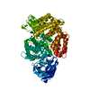


 PDBj
PDBj

 Assembly
 unidentified baculovirus


 Mass: 65.409 Da / Num. of mol.: 1 / Source method: obtained synthetically / Formula: Zn
 Mass: 134.087 Da / Num. of mol.: 2 / Source method: obtained synthetically / Formula: C4H6O5
 Mass: 62.068 Da / Num. of mol.: 18 / Source method: obtained synthetically / Formula: C2H6O2
 Mass: 332.434 Da / Num. of mol.: 1 / Source method: obtained synthetically / Formula: C20H28O4 / Feature type: SUBJECT OF INVESTIGATION
 Sample preparation
 / Beamline: ID30B / Wavelength: 0.97625 Å
 Processing