 Movie
Movie Controller


+ Open data

- Basic information
| Entry | Database: PDB / ID: 6t4d | ||||||
|---|---|---|---|---|---|---|---|
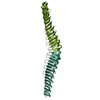
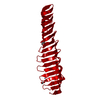
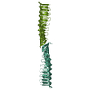
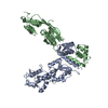
| Title | Crystal structure of Plasmodium falciparum Morn1 | ||||||
 Components Components | Morn1 | ||||||
 Keywords Keywords | STRUCTURAL PROTEIN / MORN repeat / MORN1 / Apicomplexa | ||||||
| Function / homology | Possible plasma membrane-binding motif in junctophilins, PIP-5-kinases and protein kinases. / MORN motif / MORN repeat / metal ion binding / MORN repeat-containing protein 1 / MORN repeat-containing protein 1 Function and homology information Function and homology information | ||||||
| Biological species |  | ||||||
| Method |  X-RAY DIFFRACTION / SYNCHROTRON / SAD / Resolution: 2.14 Å X-RAY DIFFRACTION / SYNCHROTRON / SAD / Resolution: 2.14 Å | ||||||
 Authors Authors | Grishkovskaya, I. / Kostan, J. / Sajko, S. / Morriswood, B. / Djinovic-Carugo, K. | ||||||
 Citation Citation | Journal: Plos One / Year: 2020 Title: Structures of three MORN repeat proteins and a re-evaluation of the proposed lipid-binding properties of MORN repeats. Authors: Sajko, S. / Grishkovskaya, I. / Kostan, J. / Graewert, M. / Setiawan, K. / Trubestein, L. / Niedermuller, K. / Gehin, C. / Sponga, A. / Puchinger, M. / Gavin, A.C. / Leonard, T.A. / Svergun, ...Authors: Sajko, S. / Grishkovskaya, I. / Kostan, J. / Graewert, M. / Setiawan, K. / Trubestein, L. / Niedermuller, K. / Gehin, C. / Sponga, A. / Puchinger, M. / Gavin, A.C. / Leonard, T.A. / Svergun, D.I. / Smith, T.K. / Morriswood, B. / Djinovic-Carugo, K. | ||||||
| History |
|
- Structure visualization
| Structure viewer | Molecule: MolmilJmol/JSmol |
|---|
- Downloads & links
-Download
| PDBx/mmCIF format | 6t4d.cif.gz | 65.1 KB | Display | PDBx/mmCIF format |
|---|---|---|---|---|
| PDB format | pdb6t4d.ent.gz | 38.2 KB | Display | PDB format |
| PDBx/mmJSON format | 6t4d.json.gz | Tree view | PDBx/mmJSON format | |
| Others |  Other downloads Other downloads |
-Validation report
| Arichive directory | https://data.pdbj.org/pub/pdb/validation_reports/t4/6t4dftp://data.pdbj.org/pub/pdb/validation_reports/t4/6t4d | HTTPS FTP |
|---|
-Related structure data


-Links
 PDBj
PDBj- Assembly
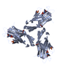
| Deposited unit | 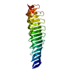
| ||||||||||||
|---|---|---|---|---|---|---|---|---|---|---|---|---|---|
| 1 | 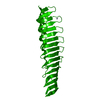
| ||||||||||||
| Unit cell |
| ||||||||||||
| Components on special symmetry positions |
|
-Components
| #1: Protein | Mass: 24717.287 Da / Num. of mol.: 1 Source method: isolated from a genetically manipulated source Source: (gene. exp.) Gene: PFFCH_03839 / Production host:  |
|---|---|
| #2: Chemical | ChemComp-ZN /   Mass: 65.409 Da / Num. of mol.: 1 / Source method: obtained synthetically / Formula: Zn Mass: 65.409 Da / Num. of mol.: 1 / Source method: obtained synthetically / Formula: Zn |
| #3: Water | ChemComp-HOH /  Mass: 18.015 Da / Num. of mol.: 12 / Source method: isolated from a natural source / Formula: H2O Mass: 18.015 Da / Num. of mol.: 12 / Source method: isolated from a natural source / Formula: H2O |
| Has ligand of interest | N |
-Experimental details
-Experiment
| Experiment | Method: X-RAY DIFFRACTION / Number of used crystals: 1 |
|---|
- Sample preparation
| Crystal | Density Matthews: 2.17 Å3/Da / Density % sol: 43.25 % |
|---|---|
| Crystal grow | Temperature: 295.15 K / Method: vapor diffusion, sitting drop / pH: 8.5 Details: 2 mM divalents mix (0.5 mM MnCl3, 0.5 mM CoCl2, 0.5 mM NiCl2, 0.5 mM Zn(OAc)2, 0.1 M Buffer System 6, pH 8.5 (Gly-Gly, AMPD), and 50% precipitation Mix 7 (20 % PEG 8000, 40% 1,5-Pentanediol) |
-Data collection
| Diffraction | Mean temperature: 100 K / Serial crystal experiment: N |
|---|---|
| Diffraction source | Source: SYNCHROTRON / Site: ESRF  / Beamline: ID29 / Wavelength: 0.976 Å / Beamline: ID29 / Wavelength: 0.976 Å |
| Detector | Type: DECTRIS PILATUS 6M / Detector: PIXEL / Date: May 12, 2016 |
| Radiation | Protocol: SINGLE WAVELENGTH / Monochromatic (M) / Laue (L): M / Scattering type: x-ray |
| Radiation wavelength | Wavelength: 0.976 Å / Relative weight: 1 |
| Reflection | Resolution: 2.14→46.43 Å / Num. obs: 12148 / % possible obs: 99.6 % / Redundancy: 4.2 % / Biso Wilson estimate: 59.97 Å2 / CC1/2: 1 / Rmerge(I) obs: 0.031 / Rpim(I) all: 0.024 / Rrim(I) all: 0.04 / Net I/σ(I): 17.5 |
| Reflection shell | Resolution: 2.14→2.2 Å / Redundancy: 3.2 % / Rmerge(I) obs: 1.065 / Mean I/σ(I) obs: 0.8 / Num. unique obs: 985 / CC1/2: 0.467 / Rpim(I) all: 0.888 / Rrim(I) all: 1.394 / % possible all: 98.1 |
- Processing
| Software |
| ||||||||||||||||||||||||||||||||||||||||||
|---|---|---|---|---|---|---|---|---|---|---|---|---|---|---|---|---|---|---|---|---|---|---|---|---|---|---|---|---|---|---|---|---|---|---|---|---|---|---|---|---|---|---|---|
| Refinement | Method to determine structure: SAD / Resolution: 2.14→41.67 Å / SU ML: 0.3556 / Cross valid method: FREE R-VALUE / σ(F): 1.34 / Phase error: 39.3704 Stereochemistry target values: GeoStd + Monomer Library + CDL v1.2
| ||||||||||||||||||||||||||||||||||||||||||
| Solvent computation | Shrinkage radii: 0.9 Å / VDW probe radii: 1.11 Å / Solvent model: FLAT BULK SOLVENT MODEL | ||||||||||||||||||||||||||||||||||||||||||
| Displacement parameters | Biso mean: 73.65 Å2 | ||||||||||||||||||||||||||||||||||||||||||
| Refinement step | Cycle: LAST / Resolution: 2.14→41.67 Å
| ||||||||||||||||||||||||||||||||||||||||||
| Refine LS restraints |
| ||||||||||||||||||||||||||||||||||||||||||
| LS refinement shell |
|
