 Movie
Movie Controller

[English] 日本語
 Yorodumi
- PDB-6szz: Crystal structure of Cold Shock Protein B (CspB) containing the m... -
+ Open data

- Basic information
| Entry | Database: PDB / ID: 6szz | ||||||
|---|---|---|---|---|---|---|---|
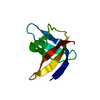
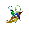
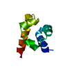
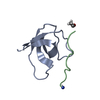
| Title | Crystal structure of Cold Shock Protein B (CspB) containing the modified residue 4-F-Trp | ||||||
 Components Components | Cold shock protein CspD | ||||||
 Keywords Keywords | STRUCTURAL PROTEIN / fluorine cold shock domain | ||||||
| Function / homology |  Function and homology information Function and homology informationregulation of RNA metabolic process / nucleoid / regulation of gene expression / nucleic acid binding / DNA binding / cytoplasm Similarity search - Function | ||||||
| Biological species |  | ||||||
| Method |  X-RAY DIFFRACTION / SYNCHROTRON / MOLECULAR REPLACEMENT / Resolution: 2.05 Å X-RAY DIFFRACTION / SYNCHROTRON / MOLECULAR REPLACEMENT / Resolution: 2.05 Å | ||||||
 Authors Authors | Zhou, T. / Mayans, O. | ||||||
 Citation Citation | Journal: Sci Rep / Year: 2020 Title: What does fluorine do to a protein? Thermodynamic, and highly-resolved structural insights into fluorine-labelled variants of the cold shock protein. Authors: Welte, H. / Zhou, T. / Mihajlenko, X. / Mayans, O. / Kovermann, M. | ||||||
| History |
|
- Structure visualization
| Structure viewer | Molecule: MolmilJmol/JSmol |
|---|
- Downloads & links
-Download
| PDBx/mmCIF format | 6szz.cif.gz | 47.2 KB | Display | PDBx/mmCIF format |
|---|---|---|---|---|
| PDB format | pdb6szz.ent.gz | 27.5 KB | Display | PDB format |
| PDBx/mmJSON format | 6szz.json.gz | Tree view | PDBx/mmJSON format | |
| Others |  Other downloads Other downloads |
-Validation report
| Arichive directory | https://data.pdbj.org/pub/pdb/validation_reports/sz/6szzftp://data.pdbj.org/pub/pdb/validation_reports/sz/6szz | HTTPS FTP |
|---|
-Related structure data
| Related structure data | 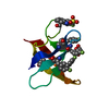 6t00C 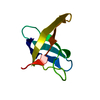 1cspS S: Starting model for refinement C: citing same article ( |
|---|---|
| Similar structure data |


-Links
 PDBj
PDBj- Assembly
| Deposited unit | 
| ||||||||||||
|---|---|---|---|---|---|---|---|---|---|---|---|---|---|
| 1 |
| ||||||||||||
| Unit cell |
|
-Components
| #1: Protein | Mass: 7390.118 Da / Num. of mol.: 1 Source method: isolated from a genetically manipulated source Source: (gene. exp.) Gene: B4122_0412, B4417_4327, CJ481_22435, DFO69_3150, ETA10_05025, ETK61_05130, ETL41_18150, FA024_03420, SC09_Contig19orf00064 Production host: | ||||
|---|---|---|---|---|---|
| #2: Chemical |   Mass: 92.094 Da / Num. of mol.: 2 / Source method: obtained synthetically / Formula: C3H8O3 Mass: 92.094 Da / Num. of mol.: 2 / Source method: obtained synthetically / Formula: C3H8O3#3: Water | ChemComp-HOH / |  Mass: 18.015 Da / Num. of mol.: 18 / Source method: isolated from a natural source / Formula: H2O Mass: 18.015 Da / Num. of mol.: 18 / Source method: isolated from a natural source / Formula: H2OHas ligand of interest | Y | |
-Experimental details
-Experiment
| Experiment | Method: X-RAY DIFFRACTION / Number of used crystals: 1 |
|---|
- Sample preparation
| Crystal | Density Matthews: 2.93 Å3/Da / Density % sol: 58.01 % |
|---|---|
| Crystal grow | Temperature: 291 K / Method: vapor diffusion, sitting drop Details: 0.1 M BIS-TRIS propane pH 7.0,1.2 M Sodium citrate tribasic dihydrate |
-Data collection
| Diffraction | Mean temperature: 100 K / Serial crystal experiment: N |
|---|---|
| Diffraction source | Source: SYNCHROTRON / Site: SLS  / Beamline: X06DA / Wavelength: 1 Å / Beamline: X06DA / Wavelength: 1 Å |
| Detector | Type: DECTRIS PILATUS 2M-F / Detector: PIXEL / Date: Jun 16, 2019 |
| Radiation | Protocol: SINGLE WAVELENGTH / Monochromatic (M) / Laue (L): M / Scattering type: x-ray |
| Radiation wavelength | Wavelength: 1 Å / Relative weight: 1 |
| Reflection | Resolution: 2.05→39.64 Å / Num. obs: 5913 / % possible obs: 99.8 % / Redundancy: 24.75 % / Biso Wilson estimate: 50.99 Å2 / CC1/2: 1 / Rsym value: 0.075 / Net I/σ(I): 28.89 |
| Reflection shell | Resolution: 2.05→2.15 Å / Num. unique obs: 762 / CC1/2: 0.584 / Rsym value: 2.735 |
- Processing
| Software |
| ||||||||||||||||||||||||||||||||||||||||
|---|---|---|---|---|---|---|---|---|---|---|---|---|---|---|---|---|---|---|---|---|---|---|---|---|---|---|---|---|---|---|---|---|---|---|---|---|---|---|---|---|---|
| Refinement | Method to determine structure: MOLECULAR REPLACEMENT Starting model: 1CSP Resolution: 2.05→39.64 Å / SU ML: 0.2461 / Cross valid method: FREE R-VALUE / σ(F): 1.35 / Phase error: 32.9561
| ||||||||||||||||||||||||||||||||||||||||
| Solvent computation | Shrinkage radii: 0.9 Å / VDW probe radii: 1.11 Å | ||||||||||||||||||||||||||||||||||||||||
| Refinement step | Cycle: LAST / Resolution: 2.05→39.64 Å
| ||||||||||||||||||||||||||||||||||||||||
| Refine LS restraints |
| ||||||||||||||||||||||||||||||||||||||||
| LS refinement shell |
| ||||||||||||||||||||||||||||||||||||||||
| Refinement TLS params. | Method: refined / Origin x: 15.596043507 Å / Origin y: 7.78346539785 Å / Origin z: 11.6198798494 Å
| ||||||||||||||||||||||||||||||||||||||||
| Refinement TLS group | Selection details: (chain 'A' and resid 1 through 67) |
