- PDB-6skd: Crystal Structure of Human Kallikrein 6 (I218Y) in complex with G... -
+
Open data
ID or keywords:
Loading...
-
Basic information
Entry
Database: PDB / ID: 6skd
Title
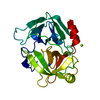
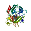
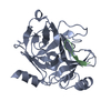
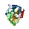
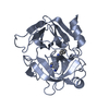
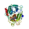
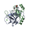
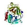
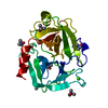
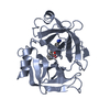
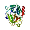
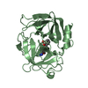
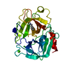
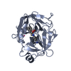
Crystal Structure of Human Kallikrein 6 (I218Y) in complex with GSK3397892A
Components
Kallikrein-6
Keywords
HYDROLASE / Protease / Inhibitor / Complex
Function / homology
Function and homology information
cornified envelope / tissue regeneration / hormone metabolic process / positive regulation of G protein-coupled receptor signaling pathway / amyloid precursor protein metabolic process / regulation of neuron projection development / protein autoprocessing / collagen catabolic process / regulation of cell differentiation / Hydrolases; Acting on peptide bonds (peptidases); Serine endopeptidases ...cornified envelope / tissue regeneration / hormone metabolic process / positive regulation of G protein-coupled receptor signaling pathway / amyloid precursor protein metabolic process / regulation of neuron projection development / protein autoprocessing / collagen catabolic process / regulation of cell differentiation / Hydrolases; Acting on peptide bonds (peptidases); Serine endopeptidases / myelination / secretory granule / central nervous system development / protein maturation / response to wounding / serine-type endopeptidase activity / nucleolus / mitochondrion / : / extracellular region / nucleoplasm / cytoplasm Similarity search - Function
Serine proteases, trypsin family, histidine active site / Serine proteases, trypsin family, serine active site / Serine proteases, trypsin family, histidine active site. / Serine proteases, trypsin family, serine active site. / Peptidase S1A, chymotrypsin family / Serine proteases, trypsin domain profile. / Trypsin-like serine protease / Serine proteases, trypsin domain / Trypsin / Trypsin-like serine proteases ...Serine proteases, trypsin family, histidine active site / Serine proteases, trypsin family, serine active site / Serine proteases, trypsin family, histidine active site. / Serine proteases, trypsin family, serine active site. / Peptidase S1A, chymotrypsin family / Serine proteases, trypsin domain profile. / Trypsin-like serine protease / Serine proteases, trypsin domain / Trypsin / Trypsin-like serine proteases / Thrombin, subunit H / Peptidase S1, PA clan, chymotrypsin-like fold / Peptidase S1, PA clan / Beta Barrel / Mainly Beta Similarity search - Domain/homology
In the structure databanks used in Yorodumi, some data are registered as the other names, "COVID-19 virus" and "2019-nCoV". Here are the details of the virus and the list of structure data.
Jan 31, 2019. EMDB accession codes are about to change! (news from PDBe EMDB page)
EMDB accession codes are about to change! (news from PDBe EMDB page)
The allocation of 4 digits for EMDB accession codes will soon come to an end. Whilst these codes will remain in use, new EMDB accession codes will include an additional digit and will expand incrementally as the available range of codes is exhausted. The current 4-digit format prefixed with “EMD-” (i.e. EMD-XXXX) will advance to a 5-digit format (i.e. EMD-XXXXX), and so on. It is currently estimated that the 4-digit codes will be depleted around Spring 2019, at which point the 5-digit format will come into force.
The EM Navigator/Yorodumi systems omit the EMD- prefix.
Related info.:Q: What is EMD? / ID/Accession-code notation in Yorodumi/EM Navigator
Yorodumi is a browser for structure data from EMDB, PDB, SASBDB, etc.
This page is also the successor to EM Navigator detail page, and also detail information page/front-end page for Omokage search.
The word "yorodu" (or yorozu) is an old Japanese word meaning "ten thousand". "mi" (miru) is to see.
Related info.:EMDB / PDB / SASBDB / Comparison of 3 databanks / Yorodumi Search / Aug 31, 2016. New EM Navigator & Yorodumi / Yorodumi Papers / Jmol/JSmol / Function and homology information / Changes in new EM Navigator and Yorodumi
 Movie
Movie Controller

 Yorodumi
 Open data
 Basic information
 Components
 Keywords
 Function and homology information
 Homo sapiens (human)
 X-RAY DIFFRACTION /
  Authors
 Citation
 Structure visualization
 Downloads & links
 Other downloads
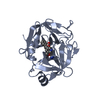


 PDBj
PDBj Assembly
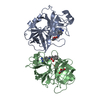

 Baculovirus expression vector pFastBac1-HM
 Num. of mol.: 2 / Source method: obtained synthetically
 Mass: 295.144 Da / Num. of mol.: 2 / Source method: obtained synthetically / Formula: C16H18BN3O2 / Feature type: SUBJECT OF INVESTIGATION
 Mass: 92.094 Da / Num. of mol.: 2 / Source method: obtained synthetically / Formula: C3H8O3
 Mass: 18.015 Da / Num. of mol.: 115 / Source method: isolated from a natural source / Formula: H2O
 Sample preparation
 / Beamline: I04 / Wavelength: 0.97949 Å
 Processing