 Movie
Movie Controller

[English] 日本語
 Yorodumi
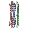
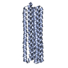
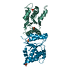
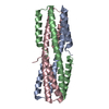
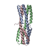
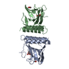
- PDB-6shk: High resolution structure of the antimicrobial peptide Dermcidin ... -
+ Open data

- Basic information
| Entry | Database: PDB / ID: 6shk | ||||||
|---|---|---|---|---|---|---|---|
| Title | High resolution structure of the antimicrobial peptide Dermcidin from human | ||||||
 Components Components | Dermcidin | ||||||
 Keywords Keywords | ANTIMICROBIAL PROTEIN / AMP / antimicrobial peptides / channel / barrel stave model | ||||||
| Function / homology |  Function and homology information Function and homology information: / Hydrolases; Acting on peptide bonds (peptidases) / Antimicrobial peptides / monoatomic ion channel activity / defense response to fungus / peptidase activity / antimicrobial humoral immune response mediated by antimicrobial peptide / defense response to bacterium / lipid binding / proteolysis ...: / Hydrolases; Acting on peptide bonds (peptidases) / Antimicrobial peptides / monoatomic ion channel activity / defense response to fungus / peptidase activity / antimicrobial humoral immune response mediated by antimicrobial peptide / defense response to bacterium / lipid binding / proteolysis / : / RNA binding / extracellular exosome / extracellular region / membrane / metal ion binding Similarity search - Function | ||||||
| Biological species |  Homo sapiens (human) Homo sapiens (human) | ||||||
| Method |  X-RAY DIFFRACTION / SYNCHROTRON / MOLECULAR REPLACEMENT / Resolution: 1.992 Å X-RAY DIFFRACTION / SYNCHROTRON / MOLECULAR REPLACEMENT / Resolution: 1.992 Å | ||||||
 Authors Authors | Zeth, K. | ||||||
 Citation Citation | Journal: To be published Title: Crystal Structure and Functional Mechanism of a Human Antimicrobial Membrane Channel Authors: Zeth, K. #1: Journal: Proc.Natl.Acad.Sci.USA / Year: 2013Title: Crystal structure and functional mechanism of a human antimicrobial membrane channel. Authors: Song, C. / Weichbrodt, C. / Salnikov, E.S. / Dynowski, M. / Forsberg, B.O. / Bechinger, B. / Steinem, C. / de Groot, B.L. / Zachariae, U. / Zeth, K. | ||||||
| History |
|
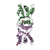
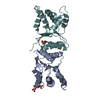
- Structure visualization
| Structure viewer | Molecule: MolmilJmol/JSmol |
|---|
- Downloads & links
-Download
| PDBx/mmCIF format | 6shk.cif.gz | 29.2 KB | Display | PDBx/mmCIF format |
|---|---|---|---|---|
| PDB format | pdb6shk.ent.gz | 19.8 KB | Display | PDB format |
| PDBx/mmJSON format | 6shk.json.gz | Tree view | PDBx/mmJSON format | |
| Others |  Other downloads Other downloads |
-Validation report
| Arichive directory | https://data.pdbj.org/pub/pdb/validation_reports/sh/6shkftp://data.pdbj.org/pub/pdb/validation_reports/sh/6shk | HTTPS FTP |
|---|
-Related structure data
| Related structure data |  2ymkSC S: Starting model for refinement C: citing same article ( |
|---|---|
| Similar structure data |


-Links
 PDBj
PDBj

- Assembly
| Deposited unit | 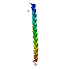
| ||||||||
|---|---|---|---|---|---|---|---|---|---|
| 1 | x 6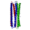
| ||||||||
| Unit cell |
|
-Components
| #1: Protein/peptide | Mass: 4826.503 Da / Num. of mol.: 1 Source method: isolated from a genetically manipulated source Source: (gene. exp.) Homo sapiens (human) / Gene: DCD, AIDD, DSEP / Production host:  References: UniProt: P81605, Hydrolases; Acting on peptide bonds (peptidases) | ||||
|---|---|---|---|---|---|
| #2: Chemical |   Mass: 65.409 Da / Num. of mol.: 2 / Source method: obtained synthetically / Formula: Zn / Feature type: SUBJECT OF INVESTIGATION Mass: 65.409 Da / Num. of mol.: 2 / Source method: obtained synthetically / Formula: Zn / Feature type: SUBJECT OF INVESTIGATIONHas ligand of interest | Y | Has protein modification | N | |
-Experimental details
-Experiment
| Experiment | Method: X-RAY DIFFRACTION / Number of used crystals: 1 |
|---|
- Sample preparation
| Crystal | Density Matthews: 2.72 Å3/Da / Density % sol: 54.77 % |
|---|---|
| Crystal grow | Temperature: 298 K / Method: vapor diffusion, sitting drop / Details: 30% PEG 4000, Ph 7, 200 mM Zinc |
-Data collection
| Diffraction | Mean temperature: 100 K / Serial crystal experiment: N |
|---|---|
| Diffraction source | Source: SYNCHROTRON / Site: PETRA III, EMBL c/o DESY  / Beamline: P13 (MX1) / Wavelength: 1 Å / Beamline: P13 (MX1) / Wavelength: 1 Å |
| Detector | Type: DECTRIS PILATUS3 S 6M / Detector: PIXEL / Date: Dec 1, 2018 |
| Radiation | Protocol: SINGLE WAVELENGTH / Monochromatic (M) / Laue (L): M / Scattering type: x-ray |
| Radiation wavelength | Wavelength: 1 Å / Relative weight: 1 |
| Reflection | Resolution: 1.99→50 Å / Num. obs: 6939 / % possible obs: 99.4 % / Redundancy: 9.34 % / CC1/2: 0.99 / Rmerge(I) obs: 0.092 / Rrim(I) all: 0.098 / Net I/σ(I): 11.32 |
| Reflection shell | Resolution: 1.99→2.11 Å / Rmerge(I) obs: 3.5 / Mean I/σ(I) obs: 0.48 / Num. unique obs: 1072 / CC1/2: 0.54 / Rrim(I) all: 3.8 |
- Processing
| Software |
| ||||||||||||||||||||||||||||||||||||||||
|---|---|---|---|---|---|---|---|---|---|---|---|---|---|---|---|---|---|---|---|---|---|---|---|---|---|---|---|---|---|---|---|---|---|---|---|---|---|---|---|---|---|
| Refinement | Method to determine structure: MOLECULAR REPLACEMENT Starting model: 2YMK Resolution: 1.992→35.293 Å / SU ML: 0.29 / Cross valid method: THROUGHOUT / σ(F): 1.33 / Phase error: 43.89
| ||||||||||||||||||||||||||||||||||||||||
| Solvent computation | Shrinkage radii: 0.9 Å / VDW probe radii: 1.11 Å | ||||||||||||||||||||||||||||||||||||||||
| Displacement parameters | Biso max: 157.98 Å2 / Biso mean: 79.3895 Å2 / Biso min: 39.72 Å2 | ||||||||||||||||||||||||||||||||||||||||
| Refinement step | Cycle: final / Resolution: 1.992→35.293 Å
| ||||||||||||||||||||||||||||||||||||||||
| LS refinement shell | Refine-ID: X-RAY DIFFRACTION / Rfactor Rfree error: 0
| ||||||||||||||||||||||||||||||||||||||||
| Refinement TLS params. | Method: refined / Origin x: 0.3332 Å / Origin y: 10.2572 Å / Origin z: 50.5063 Å
| ||||||||||||||||||||||||||||||||||||||||
| Refinement TLS group | Selection details: chain 'A' and (resid 1 through 48 ) |
