 Movie
Movie Controller

[English] 日本語
 Yorodumi
- PDB-6s7q: Crystal structure of ergothioneine degrading enzyme Ergothionase ... -
+ Open data

- Basic information
| Entry | Database: PDB / ID: 6s7q | |||||||||
|---|---|---|---|---|---|---|---|---|---|---|
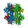
| Title | Crystal structure of ergothioneine degrading enzyme Ergothionase from Treponema denticola in complex with desmethyl-ergothioneine sulfonic acid | |||||||||
 Components Components | ergothionase | |||||||||
 Keywords Keywords | LYASE / ergothioneine degrading enzyme / TdETL / trimethlyammonium lyase / ergothioneine | |||||||||
| Function / homology | ammonia-lyase activity / Aromatic amino acid lyase / Aromatic amino acid lyase / Fumarase/histidase, N-terminal / L-Aspartase-like / Chem-KZ5 / Histidine ammonia-lyase Function and homology information Function and homology information | |||||||||
| Biological species |  Treponema denticola (bacteria) Treponema denticola (bacteria) | |||||||||
| Method |  X-RAY DIFFRACTION / SYNCHROTRON / MOLECULAR REPLACEMENT / Resolution: 2.7 Å X-RAY DIFFRACTION / SYNCHROTRON / MOLECULAR REPLACEMENT / Resolution: 2.7 Å | |||||||||
 Authors Authors | Leisinger, F. / Seebeck, F.P. | |||||||||
| Funding support |  Switzerland, 2items Switzerland, 2items
| |||||||||
 Citation Citation | Journal: Chemistry / Year: 2019 Title: Structure and Mechanism of Ergothionase from Treponema denticola. Authors: Maurer, A. / Leisinger, F. / Lim, D. / Seebeck, F.P. | |||||||||
| History |
|
- Structure visualization
| Structure viewer | Molecule: MolmilJmol/JSmol |
|---|
- Downloads & links
-Download
| PDBx/mmCIF format | 6s7q.cif.gz | 752 KB | Display | PDBx/mmCIF format |
|---|---|---|---|---|
| PDB format | pdb6s7q.ent.gz | 624.6 KB | Display | PDB format |
| PDBx/mmJSON format | 6s7q.json.gz | Tree view | PDBx/mmJSON format | |
| Others |  Other downloads Other downloads |
-Validation report
| Summary document | 6s7q_validation.pdf.gz | 2.5 MB | Display | wwPDB validaton report |
|---|---|---|---|---|
| Full document | 6s7q_full_validation.pdf.gz | 2.6 MB | Display | |
| Data in XML | 6s7q_validation.xml.gz | 143.1 KB | Display | |
| Data in CIF | 6s7q_validation.cif.gz | 189.6 KB | Display | |
| Arichive directory | https://data.pdbj.org/pub/pdb/validation_reports/s7/6s7qftp://data.pdbj.org/pub/pdb/validation_reports/s7/6s7q | HTTPS FTP |
-Related structure data
| Related structure data | 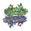 6s7jC  1gkmS S: Starting model for refinement C: citing same article ( |
|---|---|
| Similar structure data |


-Links
 PDBj
PDBj

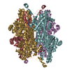
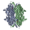
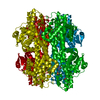
- Assembly
| Deposited unit | 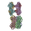
| |||||||||||||||||||||||||||||||||||||||||||||||||||||||||||||||
|---|---|---|---|---|---|---|---|---|---|---|---|---|---|---|---|---|---|---|---|---|---|---|---|---|---|---|---|---|---|---|---|---|---|---|---|---|---|---|---|---|---|---|---|---|---|---|---|---|---|---|---|---|---|---|---|---|---|---|---|---|---|---|---|---|
| 1 | 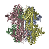
| |||||||||||||||||||||||||||||||||||||||||||||||||||||||||||||||
| 2 | 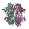
| |||||||||||||||||||||||||||||||||||||||||||||||||||||||||||||||
| Unit cell |
| |||||||||||||||||||||||||||||||||||||||||||||||||||||||||||||||
| Noncrystallographic symmetry (NCS) | NCS domain:
NCS domain segments: Component-ID: 1 / Ens-ID: 1 / Beg auth comp-ID: ASP / Beg label comp-ID: ASP / End auth comp-ID: ILE / End label comp-ID: ILE / Auth seq-ID: 2 - 498 / Label seq-ID: 1 - 497
|
-Components
| #1: Protein | Mass: 55696.668 Da / Num. of mol.: 8 Source method: isolated from a genetically manipulated source Source: (gene. exp.) Treponema denticola (bacteria) / Gene: HMPREF9733_00339 / Production host: #2: Chemical | ChemComp-KZ5 / (   Mass: 263.271 Da / Num. of mol.: 8 / Source method: obtained synthetically / Formula: C8H13N3O5S / Feature type: SUBJECT OF INVESTIGATION Mass: 263.271 Da / Num. of mol.: 8 / Source method: obtained synthetically / Formula: C8H13N3O5S / Feature type: SUBJECT OF INVESTIGATION#3: Water | ChemComp-HOH / |  Mass: 18.015 Da / Num. of mol.: 199 / Source method: isolated from a natural source / Formula: H2O Mass: 18.015 Da / Num. of mol.: 199 / Source method: isolated from a natural source / Formula: H2OHas ligand of interest | Y | |
|---|
-Experimental details
-Experiment
| Experiment | Method: X-RAY DIFFRACTION / Number of used crystals: 1 |
|---|
- Sample preparation
| Crystal | Density Matthews: 2.26 Å3/Da / Density % sol: 45.46 % |
|---|---|
| Crystal grow | Temperature: 293.5 K / Method: vapor diffusion, sitting drop / Details: PEG4000, PEG200, CaCl2, Tris-HCl |
-Data collection
| Diffraction | Mean temperature: 100 K / Serial crystal experiment: N | ||||||||||||||||||||||||||||||
|---|---|---|---|---|---|---|---|---|---|---|---|---|---|---|---|---|---|---|---|---|---|---|---|---|---|---|---|---|---|---|---|
| Diffraction source | Source: SYNCHROTRON / Site: SLS / Beamline: X06DA / Wavelength: 0.979399 Å | ||||||||||||||||||||||||||||||
| Detector | Type: DECTRIS PILATUS 2M-F / Detector: PIXEL / Date: Jun 23, 2018 | ||||||||||||||||||||||||||||||
| Radiation | Protocol: SINGLE WAVELENGTH / Monochromatic (M) / Laue (L): M / Scattering type: x-ray | ||||||||||||||||||||||||||||||
| Radiation wavelength | Wavelength: 0.979399 Å / Relative weight: 1 | ||||||||||||||||||||||||||||||
| Reflection | Resolution: 2.7→49.05 Å / Num. obs: 105530 / % possible obs: 98.7 % / Redundancy: 3.5 % / Biso Wilson estimate: 23.93 Å2 / CC1/2: 0.988 / Rmerge(I) obs: 0.124 / Rpim(I) all: 0.078 / Rrim(I) all: 0.147 / Net I/σ(I): 5.8 / Num. measured all: 365688 / Scaling rejects: 8 | ||||||||||||||||||||||||||||||
| Reflection shell | Diffraction-ID: 1
|
- Processing
| Software |
| ||||||||||||||||||||||||||||||||||||||||||||||||||||||||||||||||||||||||||||||||||||||||||||||||||||||||||||||||||||||||||||||||||||||||||||||||||||||||||||||||||||||||||||||||||||||||||
|---|---|---|---|---|---|---|---|---|---|---|---|---|---|---|---|---|---|---|---|---|---|---|---|---|---|---|---|---|---|---|---|---|---|---|---|---|---|---|---|---|---|---|---|---|---|---|---|---|---|---|---|---|---|---|---|---|---|---|---|---|---|---|---|---|---|---|---|---|---|---|---|---|---|---|---|---|---|---|---|---|---|---|---|---|---|---|---|---|---|---|---|---|---|---|---|---|---|---|---|---|---|---|---|---|---|---|---|---|---|---|---|---|---|---|---|---|---|---|---|---|---|---|---|---|---|---|---|---|---|---|---|---|---|---|---|---|---|---|---|---|---|---|---|---|---|---|---|---|---|---|---|---|---|---|---|---|---|---|---|---|---|---|---|---|---|---|---|---|---|---|---|---|---|---|---|---|---|---|---|---|---|---|---|---|---|---|---|
| Refinement | Method to determine structure: MOLECULAR REPLACEMENT Starting model: 1gkm Resolution: 2.7→49.05 Å / SU ML: 0.4 / Cross valid method: THROUGHOUT / σ(F): 1.98 / Phase error: 28.91
| ||||||||||||||||||||||||||||||||||||||||||||||||||||||||||||||||||||||||||||||||||||||||||||||||||||||||||||||||||||||||||||||||||||||||||||||||||||||||||||||||||||||||||||||||||||||||||
| Solvent computation | Shrinkage radii: 0.9 Å / VDW probe radii: 1.11 Å | ||||||||||||||||||||||||||||||||||||||||||||||||||||||||||||||||||||||||||||||||||||||||||||||||||||||||||||||||||||||||||||||||||||||||||||||||||||||||||||||||||||||||||||||||||||||||||
| Displacement parameters | Biso max: 73.78 Å2 / Biso mean: 25.367 Å2 / Biso min: 6.75 Å2 | ||||||||||||||||||||||||||||||||||||||||||||||||||||||||||||||||||||||||||||||||||||||||||||||||||||||||||||||||||||||||||||||||||||||||||||||||||||||||||||||||||||||||||||||||||||||||||
| Refinement step | Cycle: final / Resolution: 2.7→49.05 Å
| ||||||||||||||||||||||||||||||||||||||||||||||||||||||||||||||||||||||||||||||||||||||||||||||||||||||||||||||||||||||||||||||||||||||||||||||||||||||||||||||||||||||||||||||||||||||||||
| Refine LS restraints |
| ||||||||||||||||||||||||||||||||||||||||||||||||||||||||||||||||||||||||||||||||||||||||||||||||||||||||||||||||||||||||||||||||||||||||||||||||||||||||||||||||||||||||||||||||||||||||||
| Refine LS restraints NCS |
| ||||||||||||||||||||||||||||||||||||||||||||||||||||||||||||||||||||||||||||||||||||||||||||||||||||||||||||||||||||||||||||||||||||||||||||||||||||||||||||||||||||||||||||||||||||||||||
| LS refinement shell | Refine-ID: X-RAY DIFFRACTION / Rfactor Rfree error: 0
|
