BLOC-2 complex binding / AP-3 adaptor complex binding / AP-1 adaptor complex binding / phagosome acidification / positive regulation of phosphatidylcholine biosynthetic process / endosome to melanosome transport / platelet dense granule organization / positive regulation of melanin biosynthetic process / melanosome assembly / mitochondria-associated endoplasmic reticulum membrane contact site ...BLOC-2 complex binding / AP-3 adaptor complex binding / AP-1 adaptor complex binding / phagosome acidification / positive regulation of phosphatidylcholine biosynthetic process / endosome to melanosome transport / platelet dense granule organization / positive regulation of melanin biosynthetic process / melanosome assembly / mitochondria-associated endoplasmic reticulum membrane contact site / melanosome membrane / RAB geranylgeranylation / melanosome organization / RAB GEFs exchange GTP for GDP on RABs / GTP-dependent protein binding / protein localization to membrane / positive regulation of protein localization to cell periphery / small GTPase-mediated signal transduction / phagocytic vesicle / vesicle-mediated transport / endomembrane system / small monomeric GTPase / mitochondrion organization / trans-Golgi network / intracellular protein transport / phagocytic vesicle membrane / melanosome / protein transport / cell body / G protein activity / early endosome / lysosome / GTPase activity / GTP binding / perinuclear region of cytoplasm / mitochondrion / membrane / metal ion binding / plasma membrane / cytosol Similarity search - Function
Ras-related protein Rab29/Rab38/Rab32 / Small GTPase Rab domain profile. / Ran (Ras-related nuclear proteins) /TC4 subfamily of small GTPases / Rho (Ras homology) subfamily of Ras-like small GTPases / Ras subfamily of RAS small GTPases / Small GTPase / Ras family / Rab subfamily of small GTPases / Small GTP-binding protein domain / P-loop containing nucleoside triphosphate hydrolase Similarity search - Domain/homology
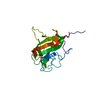
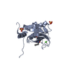
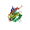
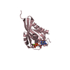
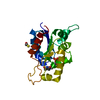
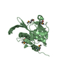
Mass: 23442.994 Da / Num. of mol.: 1 Source method: isolated from a genetically manipulated source Details: Residues not visible in the structure: 135 to 141 and 185 to end Source: (gene. exp.) Homo sapiens (human) / Gene: RAB38 / Production host: Escherichia coli (E. coli) / References: UniProt: P57729
In the structure databanks used in Yorodumi, some data are registered as the other names, "COVID-19 virus" and "2019-nCoV". Here are the details of the virus and the list of structure data.
Jan 31, 2019. EMDB accession codes are about to change! (news from PDBe EMDB page)
EMDB accession codes are about to change! (news from PDBe EMDB page)
The allocation of 4 digits for EMDB accession codes will soon come to an end. Whilst these codes will remain in use, new EMDB accession codes will include an additional digit and will expand incrementally as the available range of codes is exhausted. The current 4-digit format prefixed with “EMD-” (i.e. EMD-XXXX) will advance to a 5-digit format (i.e. EMD-XXXXX), and so on. It is currently estimated that the 4-digit codes will be depleted around Spring 2019, at which point the 5-digit format will come into force.
The EM Navigator/Yorodumi systems omit the EMD- prefix.
Related info.:Q: What is EMD? / ID/Accession-code notation in Yorodumi/EM Navigator
Yorodumi is a browser for structure data from EMDB, PDB, SASBDB, etc.
This page is also the successor to EM Navigator detail page, and also detail information page/front-end page for Omokage search.
The word "yorodu" (or yorozu) is an old Japanese word meaning "ten thousand". "mi" (miru) is to see.
Related info.:EMDB / PDB / SASBDB / Comparison of 3 databanks / Yorodumi Search / Aug 31, 2016. New EM Navigator & Yorodumi / Yorodumi Papers / Jmol/JSmol / Function and homology information / Changes in new EM Navigator and Yorodumi
 Movie
Movie Controller


 Open data
 Basic information
 Components
 Keywords
 Function and homology information
 Homo sapiens (human)
 X-RAY DIFFRACTION /
  Authors
 United Kingdom, 1items
  Citation
 Structure visualization
 Downloads & links
 Other downloads
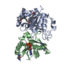


 PDBj
PDBj


 Assembly


 Mass: 523.180 Da / Num. of mol.: 1 / Source method: obtained synthetically / Formula: C10H16N5O14P3 / Feature type: SUBJECT OF INVESTIGATION / Comment: GTP, energy-carrying molecule*YM
 Mass: 62.068 Da / Num. of mol.: 9 / Source method: obtained synthetically / Formula: C2H6O2
 Mass: 24.305 Da / Num. of mol.: 1 / Source method: obtained synthetically / Formula: Mg / Feature type: SUBJECT OF INVESTIGATION
 Mass: 79.904 Da / Num. of mol.: 3 / Source method: obtained synthetically / Formula: Br
 Sample preparation
 Processing