 Movie
Movie Controller

[English] 日本語
 Yorodumi
- PDB-6s3j: Crystal Structure of lipase from Geobacillus stearothermophilus T... -
+ Open data

- Basic information
| Entry | Database: PDB / ID: 6s3j | ||||||
|---|---|---|---|---|---|---|---|
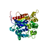
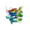
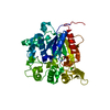
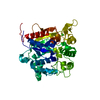
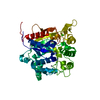
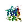
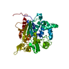
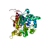
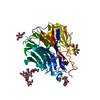
| Title | Crystal Structure of lipase from Geobacillus stearothermophilus T6 variant E134C/F149C | ||||||
 Components Components | Lipase | ||||||
 Keywords Keywords | HYDROLASE / lipase / methanol / organic solvent / disulfide bond | ||||||
| Function / homology |  Function and homology information Function and homology informationtriacylglycerol lipase / triacylglycerol lipase activity / lipid catabolic process / extracellular region / metal ion binding Similarity search - Function | ||||||
| Biological species |   Geobacillus stearothermophilus (bacteria) Geobacillus stearothermophilus (bacteria) | ||||||
| Method |  X-RAY DIFFRACTION / SYNCHROTRON / MOLECULAR REPLACEMENT / Resolution: 1.9 Å X-RAY DIFFRACTION / SYNCHROTRON / MOLECULAR REPLACEMENT / Resolution: 1.9 Å | ||||||
 Authors Authors | Gihaz, S. / Bash, Y. / Rush, I. / Shahar, A. / Pazy, Y. / Fishman, A. | ||||||
 Citation Citation | Journal: Chemcatchem / Year: 2019 Title: Bridges to Stability: Engineering Disulfide Bonds Towards Enhanced Lipase Biodiesel Synthesis Authors: Gihaz, S. / Bash, Y. / Rush, I. / Shahar, A. / Pazy, Y. / Fishman, A. | ||||||
| History |
|
- Structure visualization
| Structure viewer | Molecule: MolmilJmol/JSmol |
|---|
- Downloads & links
-Download
| PDBx/mmCIF format | 6s3j.cif.gz | 177.5 KB | Display | PDBx/mmCIF format |
|---|---|---|---|---|
| PDB format | pdb6s3j.ent.gz | 138.6 KB | Display | PDB format |
| PDBx/mmJSON format | 6s3j.json.gz | Tree view | PDBx/mmJSON format | |
| Others |  Other downloads Other downloads |
-Validation report
| Arichive directory | https://data.pdbj.org/pub/pdb/validation_reports/s3/6s3jftp://data.pdbj.org/pub/pdb/validation_reports/s3/6s3j | HTTPS FTP |
|---|
-Related structure data
| Related structure data |  6s3gC  6s3vC 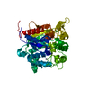 4x6uS S: Starting model for refinement C: citing same article ( |
|---|---|
| Similar structure data |

-Links
 PDBj
PDBj- Assembly
| Deposited unit | 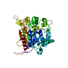
| ||||||||
|---|---|---|---|---|---|---|---|---|---|
| 1 |
| ||||||||
| Unit cell |
|
-Components
| #1: Protein | Mass: 43863.000 Da / Num. of mol.: 1 Source method: isolated from a genetically manipulated source Source: (gene. exp.) Geobacillus stearothermophilus (bacteria)Production host: |
|---|---|
| #2: Chemical | ChemComp-ZN /   Mass: 65.409 Da / Num. of mol.: 1 / Source method: obtained synthetically / Formula: Zn Mass: 65.409 Da / Num. of mol.: 1 / Source method: obtained synthetically / Formula: Zn |
| #3: Chemical | ChemComp-CA /   Mass: 40.078 Da / Num. of mol.: 1 / Source method: obtained synthetically / Formula: Ca Mass: 40.078 Da / Num. of mol.: 1 / Source method: obtained synthetically / Formula: Ca |
| #4: Water | ChemComp-HOH /  Mass: 18.015 Da / Num. of mol.: 343 / Source method: isolated from a natural source / Formula: H2O Mass: 18.015 Da / Num. of mol.: 343 / Source method: isolated from a natural source / Formula: H2O |
| Has ligand of interest | N |
| Has protein modification | Y |
-Experimental details
-Experiment
| Experiment | Method: X-RAY DIFFRACTION / Number of used crystals: 1 |
|---|
- Sample preparation
| Crystal | Density Matthews: 2.3 Å3/Da / Density % sol: 46.61 % |
|---|---|
| Crystal grow | Temperature: 293 K / Method: vapor diffusion, hanging drop / pH: 8.5 / Details: 0.2M sodium citrate, 25% PEG 3350 |
-Data collection
| Diffraction | Mean temperature: 100 K / Serial crystal experiment: N |
|---|---|
| Diffraction source | Source: SYNCHROTRON / Site: Diamond  / Beamline: I03 / Wavelength: 0.97625 Å / Beamline: I03 / Wavelength: 0.97625 Å |
| Detector | Type: DECTRIS EIGER X 16M / Detector: PIXEL / Date: Mar 6, 2019 |
| Radiation | Protocol: SINGLE WAVELENGTH / Monochromatic (M) / Laue (L): M / Scattering type: x-ray |
| Radiation wavelength | Wavelength: 0.97625 Å / Relative weight: 1 |
| Reflection | Resolution: 1.9→44.56 Å / Num. obs: 32174 / % possible obs: 98.2 % / Redundancy: 4.3 % / Biso Wilson estimate: 18.35 Å2 / CC1/2: 0.998 / Rmerge(I) obs: 0.092 / Rrim(I) all: 0.104 / Net I/σ(I): 17.5 |
| Reflection shell | Resolution: 1.9→2 Å / Redundancy: 4.8 % / Rmerge(I) obs: 0.755 / Mean I/σ(I) obs: 4.2 / Num. unique obs: 4652 / CC1/2: 0.768 / Rrim(I) all: 0.84 / % possible all: 99 |
- Processing
| Software |
| ||||||||||||||||||||
|---|---|---|---|---|---|---|---|---|---|---|---|---|---|---|---|---|---|---|---|---|---|
| Refinement | Method to determine structure: MOLECULAR REPLACEMENT Starting model: 4X6U Resolution: 1.9→44.56 Å / Cross valid method: FREE R-VALUE
| ||||||||||||||||||||
| Displacement parameters | Biso mean: 18.2 Å2 | ||||||||||||||||||||
| Refinement step | Cycle: LAST / Resolution: 1.9→44.56 Å
|
