 Movie
Movie Controller

[English] 日本語
 Yorodumi
- PDB-6s0c: Crystal structure of methionine gamma-lyase from Citrobacter freu... -
+ Open data

- Basic information
| Entry | Database: PDB / ID: 6s0c | ||||||
|---|---|---|---|---|---|---|---|
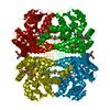
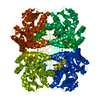
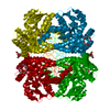
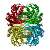
| Title | Crystal structure of methionine gamma-lyase from Citrobacter freundii modified by dimethylthiosulfinate | ||||||
 Components Components | Cystathionine gamma-synthase | ||||||
 Keywords Keywords | LYASE / METHIONINE GAMMA-LYASE | ||||||
| Function / homology |  Function and homology information Function and homology informationmethionine gamma-lyase / methionine gamma-lyase activity / transsulfuration / pyridoxal phosphate binding / cytoplasm Similarity search - Function | ||||||
| Biological species |  Citrobacter freundii (bacteria) Citrobacter freundii (bacteria) | ||||||
| Method |  X-RAY DIFFRACTION / SYNCHROTRON / MOLECULAR REPLACEMENT / Resolution: 1.46 Å X-RAY DIFFRACTION / SYNCHROTRON / MOLECULAR REPLACEMENT / Resolution: 1.46 Å | ||||||
 Authors Authors | Revtovich, S.V. / Morozova, E.A. / Nikulin, A.D. / Demidkina, T.V. | ||||||
| Funding support |  Russian Federation, 1items Russian Federation, 1items
| ||||||
 Citation Citation | Journal: Biochimie / Year: 2020 Title: Sulfoxides of sulfur-containing amino acids are suicide substrates of Citrobacter freundii methionine gamma-lyase. Structural bases of the enzyme inactivation. Authors: Revtovich, S. / Morozova, E. / Kulikova, V. / Koval, V. / Anufrieva, N. / Nikulin, A. / Demidkina, T. | ||||||
| History |
|
- Structure visualization
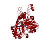
| Structure viewer | Molecule: MolmilJmol/JSmol |
|---|
- Downloads & links
-Download
| PDBx/mmCIF format | 6s0c.cif.gz | 182.7 KB | Display | PDBx/mmCIF format |
|---|---|---|---|---|
| PDB format | pdb6s0c.ent.gz | 144.8 KB | Display | PDB format |
| PDBx/mmJSON format | 6s0c.json.gz | Tree view | PDBx/mmJSON format | |
| Others |  Other downloads Other downloads |
-Validation report
| Summary document | 6s0c_validation.pdf.gz | 461.8 KB | Display | wwPDB validaton report |
|---|---|---|---|---|
| Full document | 6s0c_full_validation.pdf.gz | 464.4 KB | Display | |
| Data in XML | 6s0c_validation.xml.gz | 19.6 KB | Display | |
| Data in CIF | 6s0c_validation.cif.gz | 29.3 KB | Display | |
| Arichive directory | https://data.pdbj.org/pub/pdb/validation_reports/s0/6s0cftp://data.pdbj.org/pub/pdb/validation_reports/s0/6s0c | HTTPS FTP |
-Related structure data
| Related structure data |  5k30C  2rfvS S: Starting model for refinement C: citing same article ( |
|---|---|
| Similar structure data |


-Links
 PDBj
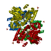
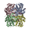
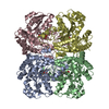
PDBj- Assembly
| Deposited unit | 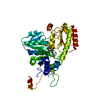
| ||||||||||||
|---|---|---|---|---|---|---|---|---|---|---|---|---|---|
| 1 | 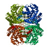
| ||||||||||||
| Unit cell |
| ||||||||||||
| Components on special symmetry positions |
|
-Components
| #1: Protein | Mass: 43075.078 Da / Num. of mol.: 1 Source method: isolated from a genetically manipulated source Source: (gene. exp.) Citrobacter freundii (bacteria)Gene: mdeA, megL, AM363_27345, AN672_11005, AZ002_001464, BED45_02240, CES93_01075, CFA70_17460, EOL10_00980, HMPREF3212_02785, NCTC13639_00292, TO64_18625 Production host: |
|---|---|
| #2: Chemical | ChemComp-PGE /   Mass: 150.173 Da / Num. of mol.: 1 / Source method: obtained synthetically / Formula: C6H14O4 Mass: 150.173 Da / Num. of mol.: 1 / Source method: obtained synthetically / Formula: C6H14O4 |
| #3: Chemical | ChemComp-PLP /   Mass: 247.142 Da / Num. of mol.: 1 / Source method: obtained synthetically / Formula: C8H10NO6P Mass: 247.142 Da / Num. of mol.: 1 / Source method: obtained synthetically / Formula: C8H10NO6P |
| #4: Water | ChemComp-HOH /  Mass: 18.015 Da / Num. of mol.: 338 / Source method: isolated from a natural source / Formula: H2O Mass: 18.015 Da / Num. of mol.: 338 / Source method: isolated from a natural source / Formula: H2O |
| Has ligand of interest | Y |
-Experimental details
-Experiment
| Experiment | Method: X-RAY DIFFRACTION / Number of used crystals: 1 |
|---|
- Sample preparation
| Crystal | Density Matthews: 2.59 Å3/Da / Density % sol: 52.56 % |
|---|---|
| Crystal grow | Temperature: 298 K / Method: vapor diffusion, hanging drop / pH: 8 / Details: 50 mM Tris-HCl, pH 8.5, 0.5 mM PLP |
-Data collection
| Diffraction | Mean temperature: 100 K / Serial crystal experiment: N |
|---|---|
| Diffraction source | Source: SYNCHROTRON / Site: BESSY  / Beamline: 14.1 / Wavelength: 0.918409 Å / Beamline: 14.1 / Wavelength: 0.918409 Å |
| Detector | Type: DECTRIS PILATUS 6M / Detector: PIXEL / Date: Jul 11, 2015 |
| Radiation | Protocol: SINGLE WAVELENGTH / Monochromatic (M) / Laue (L): M / Scattering type: x-ray |
| Radiation wavelength | Wavelength: 0.918409 Å / Relative weight: 1 |
| Reflection | Resolution: 1.46→50 Å / Num. obs: 76641 / % possible obs: 97.8 % / Redundancy: 4.45 % / CC1/2: 1 / Rrim(I) all: 0.054 / Net I/σ(I): 20.48 |
| Reflection shell | Resolution: 1.46→1.55 Å / Redundancy: 4.1 % / Mean I/σ(I) obs: 2.22 / Num. unique obs: 120129 / CC1/2: 0.75 / Rrim(I) all: 0.701 / % possible all: 70.5 |
- Processing
| Software |
| ||||||||||||||||||||||||||||||||||||||||||||||||||||||||||||||||||||||||||||||||||||||||||||||||||||||||||||||||
|---|---|---|---|---|---|---|---|---|---|---|---|---|---|---|---|---|---|---|---|---|---|---|---|---|---|---|---|---|---|---|---|---|---|---|---|---|---|---|---|---|---|---|---|---|---|---|---|---|---|---|---|---|---|---|---|---|---|---|---|---|---|---|---|---|---|---|---|---|---|---|---|---|---|---|---|---|---|---|---|---|---|---|---|---|---|---|---|---|---|---|---|---|---|---|---|---|---|---|---|---|---|---|---|---|---|---|---|---|---|---|---|---|---|
| Refinement | Method to determine structure: MOLECULAR REPLACEMENT Starting model: 2RFV Resolution: 1.46→40.411 Å / SU ML: 0.16 / Cross valid method: THROUGHOUT / σ(F): 1.35 / Phase error: 17.97
| ||||||||||||||||||||||||||||||||||||||||||||||||||||||||||||||||||||||||||||||||||||||||||||||||||||||||||||||||
| Solvent computation | Shrinkage radii: 0.9 Å / VDW probe radii: 1.11 Å | ||||||||||||||||||||||||||||||||||||||||||||||||||||||||||||||||||||||||||||||||||||||||||||||||||||||||||||||||
| Refinement step | Cycle: LAST / Resolution: 1.46→40.411 Å
| ||||||||||||||||||||||||||||||||||||||||||||||||||||||||||||||||||||||||||||||||||||||||||||||||||||||||||||||||
| Refine LS restraints |
| ||||||||||||||||||||||||||||||||||||||||||||||||||||||||||||||||||||||||||||||||||||||||||||||||||||||||||||||||
| LS refinement shell |
|
