[English] 日本語
 Yorodumi
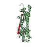
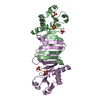
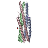
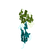
- PDB-6rx3: Crystal structure of human syncytin 2 in post-fusion conformation -
+ Open data
- Basic information
| Entry | Database: PDB / ID: 6rx3 | ||||||
|---|---|---|---|---|---|---|---|
| Title | Crystal structure of human syncytin 2 in post-fusion conformation | ||||||
 Components Components | Syncytin-2 | ||||||
 Keywords Keywords | MEMBRANE PROTEIN / HUMAN PLACENTAL PROTEIN / MEMBRANE FUSION / ENDOGENOUS RETROVIRUS / HERV-FRD / SYNCYTIN | ||||||
| Function / homology |  Function and homology information Function and homology informationsyncytium formation / syncytium formation by plasma membrane fusion / myoblast fusion / plasma membrane Similarity search - Function | ||||||
| Biological species |  Homo sapiens (human) Homo sapiens (human) | ||||||
| Method |  X-RAY DIFFRACTION / X-RAY DIFFRACTION /  SYNCHROTRON / SYNCHROTRON /  MOLECULAR REPLACEMENT / Resolution: 2.2 Å MOLECULAR REPLACEMENT / Resolution: 2.2 Å | ||||||
 Authors Authors | Ruigrok, K. / Backovic, M. / Vaney, M.C. / Rey, F.A. | ||||||
| Funding support |  France, 1items France, 1items
| ||||||
 Citation Citation |  Journal: J.Mol.Biol. / Year: 2019 Journal: J.Mol.Biol. / Year: 2019Title: X-ray Structures of the Post-fusion 6-Helix Bundle of the Human Syncytins and their Functional Implications. Authors: Ruigrok, K. / Vaney, M.C. / Buchrieser, J. / Baquero, E. / Hellert, J. / Baron, B. / England, P. / Schwartz, O. / Rey, F.A. / Backovic, M. #1:  Journal: J.Mol.Biol. / Year: 2005 Journal: J.Mol.Biol. / Year: 2005Title: Crystal structure of a pivotal domain of human syncytin-2, a 40 million years old endogenous retrovirus fusogenic envelope gene captured by primates. Authors: Renard, M. / Varela, P.F. / Letzelter, C. / Duquerroy, S. / Rey, F.A. / Heidmann, T. | ||||||
| History |
|
- Structure visualization
| Structure viewer | Molecule:  Molmil Molmil Jmol/JSmol Jmol/JSmol |
|---|
- Downloads & links
- Download
| PDBx/mmCIF format |  6rx3.cif.gz 6rx3.cif.gz | 69.4 KB | Display |  PDBx/mmCIF format PDBx/mmCIF format |
|---|---|---|---|---|
| PDB format |  pdb6rx3.ent.gz pdb6rx3.ent.gz | 50.8 KB | Display |  PDB format PDB format |
| PDBx/mmJSON format |  6rx3.json.gz 6rx3.json.gz | Tree view |  PDBx/mmJSON format PDBx/mmJSON format | |
| Others |  Other downloads Other downloads |
-Validation report
| Summary document |  6rx3_validation.pdf.gz 6rx3_validation.pdf.gz | 437.1 KB | Display |  wwPDB validaton report wwPDB validaton report |
|---|---|---|---|---|
| Full document |  6rx3_full_validation.pdf.gz 6rx3_full_validation.pdf.gz | 437.3 KB | Display | |
| Data in XML |  6rx3_validation.xml.gz 6rx3_validation.xml.gz | 12.3 KB | Display | |
| Data in CIF |  6rx3_validation.cif.gz 6rx3_validation.cif.gz | 17 KB | Display | |
| Arichive directory |  https://data.pdbj.org/pub/pdb/validation_reports/rx/6rx3 https://data.pdbj.org/pub/pdb/validation_reports/rx/6rx3 ftp://data.pdbj.org/pub/pdb/validation_reports/rx/6rx3 ftp://data.pdbj.org/pub/pdb/validation_reports/rx/6rx3 | HTTPS FTP |
-Related structure data
| Related structure data |  6rx1SC S: Starting model for refinement C: citing same article ( |
|---|---|
| Similar structure data |
- Links
Links
- Assembly
| Deposited unit | 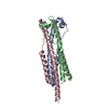
| ||||||||
|---|---|---|---|---|---|---|---|---|---|
| 1 |
| ||||||||
| Unit cell |
|
- Components
| #1: Protein | Mass: 12196.797 Da / Num. of mol.: 3 Source method: isolated from a genetically manipulated source Details: N-ter His-tag:MHHHHHH TEV cleavage site: ENLYFQS / Source: (gene. exp.)  Homo sapiens (human) / Gene: ERVFRD-1, ERVFRDE1, UNQ6191/PRO20218 / Plasmid: pET28a / Production host: Homo sapiens (human) / Gene: ERVFRD-1, ERVFRDE1, UNQ6191/PRO20218 / Plasmid: pET28a / Production host:  #2: Chemical | ChemComp-CL / | #3: Water | ChemComp-HOH / | Has protein modification | Y | |
|---|
-Experimental details
-Experiment
| Experiment | Method:  X-RAY DIFFRACTION / Number of used crystals: 1 X-RAY DIFFRACTION / Number of used crystals: 1 |
|---|
- Sample preparation
| Crystal | Density Matthews: 2.3 Å3/Da / Density % sol: 46.6 % |
|---|---|
| Crystal grow | Temperature: 291 K / Method: vapor diffusion, sitting drop / pH: 8.5 / Details: 0.1 M Tris pH 8.5, 25% v/v tert-butanol |
-Data collection
| Diffraction | Mean temperature: 100 K / Serial crystal experiment: N |
|---|---|
| Diffraction source | Source:  SYNCHROTRON / Site: SYNCHROTRON / Site:  SOLEIL SOLEIL  / Beamline: PROXIMA 2 / Wavelength: 0.98 Å / Beamline: PROXIMA 2 / Wavelength: 0.98 Å |
| Detector | Type: DECTRIS EIGER X 9M / Detector: PIXEL / Date: Nov 10, 2017 |
| Radiation | Monochromator: cryogenically cooled channel-cut Si[111] / Protocol: SINGLE WAVELENGTH / Monochromatic (M) / Laue (L): M / Scattering type: x-ray |
| Radiation wavelength | Wavelength: 0.98 Å / Relative weight: 1 |
| Reflection | Resolution: 2.2→45.7 Å / Num. obs: 17310 / % possible obs: 99.5 % / Redundancy: 11.3 % / Biso Wilson estimate: 57.03 Å2 / CC1/2: 1 / Rmerge(I) obs: 0.118 / Rpim(I) all: 0.053 / Rrim(I) all: 0.13 / Net I/σ(I): 11.5 |
| Reflection shell | Resolution: 2.2→2.27 Å / Redundancy: 11.5 % / Rmerge(I) obs: 0.1977 / Mean I/σ(I) obs: 1.1 / Num. unique obs: 1474 / CC1/2: 0.527 / Rpim(I) all: 0.873 / Rrim(I) all: 0.2164 / % possible all: 99 |
- Processing
| Software |
| ||||||||||||||||||||||||||||||||||||||||||||||||||||||||||||||||||||||||||||||||||||||||||||||||||||||||||||||||||
|---|---|---|---|---|---|---|---|---|---|---|---|---|---|---|---|---|---|---|---|---|---|---|---|---|---|---|---|---|---|---|---|---|---|---|---|---|---|---|---|---|---|---|---|---|---|---|---|---|---|---|---|---|---|---|---|---|---|---|---|---|---|---|---|---|---|---|---|---|---|---|---|---|---|---|---|---|---|---|---|---|---|---|---|---|---|---|---|---|---|---|---|---|---|---|---|---|---|---|---|---|---|---|---|---|---|---|---|---|---|---|---|---|---|---|---|
| Refinement | Method to determine structure:  MOLECULAR REPLACEMENT MOLECULAR REPLACEMENTStarting model: 6RX1 Resolution: 2.2→45.7 Å / Cor.coef. Fo:Fc: 0.941 / Cor.coef. Fo:Fc free: 0.939 / SU R Cruickshank DPI: 0.243 / Cross valid method: THROUGHOUT / σ(F): 0 / SU R Blow DPI: 0.254 / SU Rfree Blow DPI: 0.205 / SU Rfree Cruickshank DPI: 0.202
| ||||||||||||||||||||||||||||||||||||||||||||||||||||||||||||||||||||||||||||||||||||||||||||||||||||||||||||||||||
| Displacement parameters | Biso mean: 59.58 Å2
| ||||||||||||||||||||||||||||||||||||||||||||||||||||||||||||||||||||||||||||||||||||||||||||||||||||||||||||||||||
| Refine analyze | Luzzati coordinate error obs: 0.31 Å | ||||||||||||||||||||||||||||||||||||||||||||||||||||||||||||||||||||||||||||||||||||||||||||||||||||||||||||||||||
| Refinement step | Cycle: 1 / Resolution: 2.2→45.7 Å
| ||||||||||||||||||||||||||||||||||||||||||||||||||||||||||||||||||||||||||||||||||||||||||||||||||||||||||||||||||
| Refine LS restraints |
| ||||||||||||||||||||||||||||||||||||||||||||||||||||||||||||||||||||||||||||||||||||||||||||||||||||||||||||||||||
| LS refinement shell | Resolution: 2.2→2.33 Å / Total num. of bins used: 9
|
 Movie
Movie Controller




 PDBj
PDBj


