 Movie
Movie Controller

[English] 日本語
 Yorodumi
- PDB-6rl1: The crystal structure of AbnE, an arabino-oligosaccharide binding... -
+ Open data

- Basic information
| Entry | Database: PDB / ID: 6rl1 | |||||||||
|---|---|---|---|---|---|---|---|---|---|---|
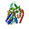
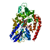
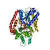
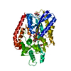
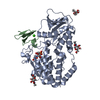
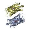
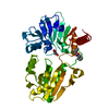
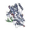
| Title | The crystal structure of AbnE, an arabino-oligosaccharide binding protein, in complex with arabinotriose | |||||||||
 Components Components | Arabino-oligosaccharids-binding protein | |||||||||
 Keywords Keywords | SUGAR BINDING PROTEIN / ABC transporter / Geobacillus stearothermophilus / arabinotriose / arabino-oligosaccharide | |||||||||
| Function / homology | : / Bacterial extracellular solute-binding protein / Bacterial extracellular solute-binding protein / Prokaryotic membrane lipoprotein lipid attachment site profile. / metal ion binding / Arabino-oligosaccharids-binding protein Function and homology information Function and homology information | |||||||||
| Biological species |   Geobacillus stearothermophilus (bacteria) Geobacillus stearothermophilus (bacteria) | |||||||||
| Method |  X-RAY DIFFRACTION / SYNCHROTRON / MOLECULAR REPLACEMENT / Resolution: 1.7 Å X-RAY DIFFRACTION / SYNCHROTRON / MOLECULAR REPLACEMENT / Resolution: 1.7 Å | |||||||||
 Authors Authors | Lansky, S. / Salama, R. / Shoham, Y. / Shoham, G. | |||||||||
 Citation Citation | Journal: J.Mol.Biol. / Year: 2020 Title: Carbohydrate-Binding Capability and Functional Conformational Changes of AbnE, an Arabino-oligosaccharide Binding Protein. Authors: Lansky, S. / Salama, R. / Shulami, S. / Lavid, N. / Sen, S. / Schapiro, I. / Shoham, Y. / Shoham, G. | |||||||||
| History |
|
- Structure visualization
| Structure viewer | Molecule: MolmilJmol/JSmol |
|---|
- Downloads & links
-Download
| PDBx/mmCIF format | 6rl1.cif.gz | 118.2 KB | Display | PDBx/mmCIF format |
|---|---|---|---|---|
| PDB format | pdb6rl1.ent.gz | 86 KB | Display | PDB format |
| PDBx/mmJSON format | 6rl1.json.gz | Tree view | PDBx/mmJSON format | |
| Others |  Other downloads Other downloads |
-Validation report
| Arichive directory | https://data.pdbj.org/pub/pdb/validation_reports/rl/6rl1ftp://data.pdbj.org/pub/pdb/validation_reports/rl/6rl1 | HTTPS FTP |
|---|
-Related structure data
| Related structure data |  6rjyC  6rkhSC  6rkjC  6rklC  6rkxC 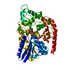 6rl2C C: citing same article ( S: Starting model for refinement |
|---|---|
| Similar structure data |


-Links
 PDBj
PDBj


- Assembly
| Deposited unit | 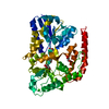
| ||||||||
|---|---|---|---|---|---|---|---|---|---|
| 1 |
| ||||||||
| Unit cell |
|
-Components
| #1: Protein | Mass: 48689.836 Da / Num. of mol.: 1 Source method: isolated from a genetically manipulated source Source: (gene. exp.) Geobacillus stearothermophilus (bacteria)Gene: abnE / Production host: | ||||
|---|---|---|---|---|---|
| #2: Polysaccharide | alpha-L-arabinofuranose-(1-5)-alpha-L-arabinofuranose-(1-5)-alpha-L-arabinofuranose Source method: isolated from a genetically manipulated source | ||||
| #3: Chemical |   Mass: 92.094 Da / Num. of mol.: 3 / Source method: obtained synthetically / Formula: C3H8O3 Mass: 92.094 Da / Num. of mol.: 3 / Source method: obtained synthetically / Formula: C3H8O3#4: Chemical | ChemComp-CA / |   Mass: 40.078 Da / Num. of mol.: 1 / Source method: obtained synthetically / Formula: Ca Mass: 40.078 Da / Num. of mol.: 1 / Source method: obtained synthetically / Formula: Ca#5: Water | ChemComp-HOH / |  Mass: 18.015 Da / Num. of mol.: 657 / Source method: isolated from a natural source / Formula: H2O Mass: 18.015 Da / Num. of mol.: 657 / Source method: isolated from a natural source / Formula: H2O |
-Experimental details
-Experiment
| Experiment | Method: X-RAY DIFFRACTION / Number of used crystals: 1 |
|---|
- Sample preparation
| Crystal | Density Matthews: 2.41 Å3/Da / Density % sol: 49.01 % |
|---|---|
| Crystal grow | Temperature: 293 K / Method: vapor diffusion, hanging drop / pH: 9 / Details: 22% PEG 4K, 0.1M Tris/HCl buffer, pH 9 |
-Data collection
| Diffraction | Mean temperature: 100 K / Serial crystal experiment: N |
|---|---|
| Diffraction source | Source: SYNCHROTRON / Site: ESRF  / Beamline: BM30A / Wavelength: 0.98 Å / Beamline: BM30A / Wavelength: 0.98 Å |
| Detector | Type: ADSC QUANTUM 315r / Detector: CCD / Date: Feb 15, 2016 |
| Radiation | Protocol: SINGLE WAVELENGTH / Monochromatic (M) / Laue (L): M / Scattering type: x-ray |
| Radiation wavelength | Wavelength: 0.98 Å / Relative weight: 1 |
| Reflection | Resolution: 1.7→48.04 Å / Num. obs: 54441 / % possible obs: 97.7 % / Redundancy: 13.7 % / Rmerge(I) obs: 0.064 / Net I/σ(I): 29.5 |
| Reflection shell | Resolution: 1.7→1.8 Å / Redundancy: 12.7 % / Rmerge(I) obs: 0.427 / Mean I/σ(I) obs: 5.5 / Num. unique obs: 7085 / % possible all: 86 |
- Processing
| Software |
| ||||||||||||||||||||||||||||||||||||||||||||||||||||||||||||||||||||||||||||||||||||||||||||||||||||||||||||||||
|---|---|---|---|---|---|---|---|---|---|---|---|---|---|---|---|---|---|---|---|---|---|---|---|---|---|---|---|---|---|---|---|---|---|---|---|---|---|---|---|---|---|---|---|---|---|---|---|---|---|---|---|---|---|---|---|---|---|---|---|---|---|---|---|---|---|---|---|---|---|---|---|---|---|---|---|---|---|---|---|---|---|---|---|---|---|---|---|---|---|---|---|---|---|---|---|---|---|---|---|---|---|---|---|---|---|---|---|---|---|---|---|---|---|
| Refinement | Method to determine structure: MOLECULAR REPLACEMENT Starting model: 6RKH Resolution: 1.7→44.614 Å / SU ML: 0.17 / Cross valid method: FREE R-VALUE / σ(F): 1.36 / Phase error: 19.52
| ||||||||||||||||||||||||||||||||||||||||||||||||||||||||||||||||||||||||||||||||||||||||||||||||||||||||||||||||
| Solvent computation | Shrinkage radii: 0.9 Å / VDW probe radii: 1.11 Å | ||||||||||||||||||||||||||||||||||||||||||||||||||||||||||||||||||||||||||||||||||||||||||||||||||||||||||||||||
| Refinement step | Cycle: LAST / Resolution: 1.7→44.614 Å
| ||||||||||||||||||||||||||||||||||||||||||||||||||||||||||||||||||||||||||||||||||||||||||||||||||||||||||||||||
| Refine LS restraints |
| ||||||||||||||||||||||||||||||||||||||||||||||||||||||||||||||||||||||||||||||||||||||||||||||||||||||||||||||||
| LS refinement shell |
|
