 Movie
Movie Controller


+ Open data

- Basic information
| Entry | Database: PDB / ID: 6rjn | ||||||||||||
|---|---|---|---|---|---|---|---|---|---|---|---|---|---|
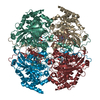
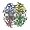
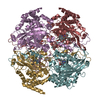
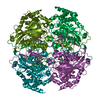
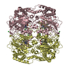
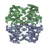
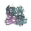
| Title | Crystal structure of a Fungal Catalase at 2.3 Angstroms | ||||||||||||
 Components Components | Catalase | ||||||||||||
 Keywords Keywords | OXIDOREDUCTASE / Pichia pastoris / Catalase / Heme / NADP | ||||||||||||
| Function / homology |  Function and homology information Function and homology informationcatalase / catalase activity / hydrogen peroxide catabolic process / response to hydrogen peroxide / peroxisome / nucleotide binding / heme binding / mitochondrion / metal ion binding Similarity search - Function | ||||||||||||
| Biological species |  Komagataella pastoris (fungus) Komagataella pastoris (fungus) | ||||||||||||
| Method |  X-RAY DIFFRACTION / SYNCHROTRON / MOLECULAR REPLACEMENT / Resolution: 2.295 Å X-RAY DIFFRACTION / SYNCHROTRON / MOLECULAR REPLACEMENT / Resolution: 2.295 Å | ||||||||||||
 Authors Authors | Gomez, S. / Navas-Yuste, S. / Payne, A.M. / Rivera, W. / Lopez-Estepa, M. / Brangbour, C. / Fulla, D. / Juanhuix, J. / Fernandez, F.J. / Vega, M.C. | ||||||||||||
| Funding support |  Spain, 3items Spain, 3items
| ||||||||||||
 Citation Citation | Journal: Free Radic. Biol. Med. / Year: 2019 Title: Peroxisomal catalases from the yeasts Pichia pastoris and Kluyveromyces lactis as models for oxidative damage in higher eukaryotes. Authors: Gomez, S. / Navas-Yuste, S. / Payne, A.M. / Rivera, W. / Lopez-Estepa, M. / Brangbour, C. / Fulla, D. / Juanhuix, J. / Fernandez, F.J. / Vega, M.C. #1: Journal: Front Microbiol / Year: 2017Title: Crystal Structure of Glyceraldehyde-3-Phosphate Dehydrogenase from the Gram-Positive Bacterial Pathogen Authors: Querol-Garcia, J. / Fernandez, F.J. / Marin, A.V. / Gomez, S. / Fulla, D. / Melchor-Tafur, C. / Franco-Hidalgo, V. / Alberti, S. / Juanhuix, J. / Rodriguez de Cordoba, S. / Regueiro, J.R. / Vega, M.C. | ||||||||||||
| History |
|
- Structure visualization
| Structure viewer | Molecule: MolmilJmol/JSmol |
|---|
- Downloads & links
-Download
| PDBx/mmCIF format | 6rjn.cif.gz | 446.7 KB | Display | PDBx/mmCIF format |
|---|---|---|---|---|
| PDB format | pdb6rjn.ent.gz | 364.5 KB | Display | PDB format |
| PDBx/mmJSON format | 6rjn.json.gz | Tree view | PDBx/mmJSON format | |
| Others |  Other downloads Other downloads |
-Validation report
| Arichive directory | https://data.pdbj.org/pub/pdb/validation_reports/rj/6rjnftp://data.pdbj.org/pub/pdb/validation_reports/rj/6rjn | HTTPS FTP |
|---|
-Related structure data
| Related structure data |  6rjrC  2xq1S S: Starting model for refinement C: citing same article ( |
|---|---|
| Similar structure data |

-Links
 PDBj
PDBj

- Assembly
| Deposited unit | 
| ||||||||
|---|---|---|---|---|---|---|---|---|---|
| 1 |
| ||||||||
| Unit cell |
|
-Components
-Protein , 1 types, 4 molecules ABCD
| #1: Protein | Mass: 58065.570 Da / Num. of mol.: 4 Source method: isolated from a genetically manipulated source Source: (gene. exp.) Komagataella pastoris (fungus) / Gene: CTA1 / Plasmid: pETM-11 / Production host:  |
|---|
-Non-polymers , 9 types, 1273 molecules 


| #2: Chemical | ChemComp-HEM /  Mass: 616.487 Da / Num. of mol.: 4 / Source method: obtained synthetically / Formula: C34H32FeN4O4 Mass: 616.487 Da / Num. of mol.: 4 / Source method: obtained synthetically / Formula: C34H32FeN4O4#3: Chemical | ChemComp-NDP /  Mass: 745.421 Da / Num. of mol.: 4 / Source method: obtained synthetically / Formula: C21H30N7O17P3 Mass: 745.421 Da / Num. of mol.: 4 / Source method: obtained synthetically / Formula: C21H30N7O17P3#4: Chemical | ChemComp-GOL /  Mass: 92.094 Da / Num. of mol.: 17 / Source method: obtained synthetically / Formula: C3H8O3 Mass: 92.094 Da / Num. of mol.: 17 / Source method: obtained synthetically / Formula: C3H8O3#5: Chemical |  Mass: 96.063 Da / Num. of mol.: 2 / Source method: obtained synthetically / Formula: SO4 Mass: 96.063 Da / Num. of mol.: 2 / Source method: obtained synthetically / Formula: SO4#6: Chemical | ChemComp-CL /  Mass: 35.453 Da / Num. of mol.: 5 / Source method: obtained synthetically / Formula: Cl Mass: 35.453 Da / Num. of mol.: 5 / Source method: obtained synthetically / Formula: Cl#7: Chemical | ChemComp-K /  Mass: 39.098 Da / Num. of mol.: 7 / Source method: obtained synthetically / Formula: K Mass: 39.098 Da / Num. of mol.: 7 / Source method: obtained synthetically / Formula: K#8: Chemical | ChemComp-NA /  Mass: 22.990 Da / Num. of mol.: 4 / Source method: obtained synthetically / Formula: Na Mass: 22.990 Da / Num. of mol.: 4 / Source method: obtained synthetically / Formula: Na#9: Chemical |  Mass: 106.120 Da / Num. of mol.: 2 / Source method: obtained synthetically / Formula: C4H10O3 Mass: 106.120 Da / Num. of mol.: 2 / Source method: obtained synthetically / Formula: C4H10O3#10: Water | ChemComp-HOH / | Mass: 18.015 Da / Num. of mol.: 1228 / Source method: isolated from a natural source / Formula: H2O |
|---|
-Experimental details
-Experiment
| Experiment | Method: X-RAY DIFFRACTION / Number of used crystals: 1 |
|---|
- Sample preparation
| Crystal | Density Matthews: 3 Å3/Da / Density % sol: 59.04 % / Description: Long thin needles |
|---|---|
| Crystal grow | Temperature: 293 K / Method: vapor diffusion, sitting drop / pH: 6.5 Details: 0.2 M sodium sulfate, 0.1 M Bis-Tris propane, pH 6.5, 20% (w/v) polyethylene glycol (PEG) 3350 |
-Data collection
| Diffraction | Mean temperature: 100 K / Serial crystal experiment: N |
|---|---|
| Diffraction source | Source: SYNCHROTRON / Site: SOLEIL  / Beamline: PROXIMA 2 / Wavelength: 0.9801 Å / Beamline: PROXIMA 2 / Wavelength: 0.9801 Å |
| Detector | Type: ADSC QUANTUM 315r / Detector: CCD / Date: Oct 26, 2015 Details: CONVEX PREFOCUSSING MIRROR AND A KIRKPATRICK-BAEZ PAIR OF FOCUSSING MIRRORS |
| Radiation | Monochromator: CRYOGENICALLY COOLED CHANNEL CUT SI[111] CRYSTAL MONOCHROMATOR Protocol: SINGLE WAVELENGTH / Monochromatic (M) / Laue (L): M / Scattering type: x-ray |
| Radiation wavelength | Wavelength: 0.9801 Å / Relative weight: 1 |
| Reflection | Resolution: 2.29→57.9 Å / Num. obs: 124735 / % possible obs: 99.5 % / Redundancy: 6.7 % / CC1/2: 0.988 / Rmerge(I) obs: 0.253 / Rpim(I) all: 0.147 / Rrim(I) all: 0.279 / Net I/σ(I): 9 |
| Reflection shell | Resolution: 2.29→2.42 Å / Redundancy: 6.6 % / Rmerge(I) obs: 1.477 / Num. unique obs: 17551 / CC1/2: 0.579 / Rpim(I) all: 0.619 / Rrim(I) all: 1.604 / % possible all: 97 |
- Processing
| Software |
| |||||||||||||||||||||||||||||||||||||||||||||||||||||||||||||||||||||||||||||||||||||||||||||||||||||||||||||||||||||||||||||||||||||||||||||||||||||||||||||||||||||||||||||||||||||||||||||||||||||||||||||||||||||||||
|---|---|---|---|---|---|---|---|---|---|---|---|---|---|---|---|---|---|---|---|---|---|---|---|---|---|---|---|---|---|---|---|---|---|---|---|---|---|---|---|---|---|---|---|---|---|---|---|---|---|---|---|---|---|---|---|---|---|---|---|---|---|---|---|---|---|---|---|---|---|---|---|---|---|---|---|---|---|---|---|---|---|---|---|---|---|---|---|---|---|---|---|---|---|---|---|---|---|---|---|---|---|---|---|---|---|---|---|---|---|---|---|---|---|---|---|---|---|---|---|---|---|---|---|---|---|---|---|---|---|---|---|---|---|---|---|---|---|---|---|---|---|---|---|---|---|---|---|---|---|---|---|---|---|---|---|---|---|---|---|---|---|---|---|---|---|---|---|---|---|---|---|---|---|---|---|---|---|---|---|---|---|---|---|---|---|---|---|---|---|---|---|---|---|---|---|---|---|---|---|---|---|---|---|---|---|---|---|---|---|---|---|---|---|---|---|---|---|---|
| Refinement | Method to determine structure: MOLECULAR REPLACEMENT Starting model: wwPDB 2XQ1 Resolution: 2.295→54.665 Å / SU ML: 0.25 / Cross valid method: FREE R-VALUE / σ(F): 1.33 / Phase error: 18.53
| |||||||||||||||||||||||||||||||||||||||||||||||||||||||||||||||||||||||||||||||||||||||||||||||||||||||||||||||||||||||||||||||||||||||||||||||||||||||||||||||||||||||||||||||||||||||||||||||||||||||||||||||||||||||||
| Solvent computation | Shrinkage radii: 0.9 Å / VDW probe radii: 1.11 Å | |||||||||||||||||||||||||||||||||||||||||||||||||||||||||||||||||||||||||||||||||||||||||||||||||||||||||||||||||||||||||||||||||||||||||||||||||||||||||||||||||||||||||||||||||||||||||||||||||||||||||||||||||||||||||
| Refinement step | Cycle: LAST / Resolution: 2.295→54.665 Å
| |||||||||||||||||||||||||||||||||||||||||||||||||||||||||||||||||||||||||||||||||||||||||||||||||||||||||||||||||||||||||||||||||||||||||||||||||||||||||||||||||||||||||||||||||||||||||||||||||||||||||||||||||||||||||
| Refine LS restraints |
| |||||||||||||||||||||||||||||||||||||||||||||||||||||||||||||||||||||||||||||||||||||||||||||||||||||||||||||||||||||||||||||||||||||||||||||||||||||||||||||||||||||||||||||||||||||||||||||||||||||||||||||||||||||||||
| LS refinement shell |
|
