 Movie
Movie Controller

[English] 日本語
 Yorodumi
- PDB-6rd8: CryoEM structure of Polytomella F-ATP synthase, c-ring position 2... -
+ Open data

- Basic information
| Entry | Database: PDB / ID: 6rd8 | |||||||||
|---|---|---|---|---|---|---|---|---|---|---|
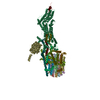
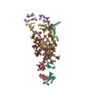
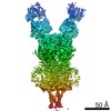
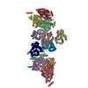
| Title | CryoEM structure of Polytomella F-ATP synthase, c-ring position 2, focussed refinement of Fo and peripheral stalk | |||||||||
 Components Components |
| |||||||||
 Keywords Keywords | PROTON TRANSPORT / mitochondrial ATP synthase dimer flexible coupling cryoEM | |||||||||
| Function / homology |  Function and homology information Function and homology informationproton transmembrane transporter activity / proton motive force-driven ATP synthesis / proton-transporting two-sector ATPase complex, proton-transporting domain / proton-transporting ATP synthase complex / proton-transporting ATP synthase activity, rotational mechanism / lipid binding / mitochondrion Similarity search - Function | |||||||||
| Biological species |  Polytomella sp. Pringsheim 198.80 (plant) Polytomella sp. Pringsheim 198.80 (plant) | |||||||||
| Method | ELECTRON MICROSCOPY / single particle reconstruction / cryo EM / Resolution: 3.08 Å | |||||||||
 Authors Authors | Murphy, B.J. / Klusch, N. / Yildiz, O. / Kuhlbrandt, W. | |||||||||
| Funding support |  Germany, 2items Germany, 2items
| |||||||||
 Citation Citation | Journal: Science / Year: 2019 Title: Rotary substates of mitochondrial ATP synthase reveal the basis of flexible F-F coupling. Authors: Bonnie J Murphy / Niklas Klusch / Julian Langer / Deryck J Mills / Özkan Yildiz / Werner Kühlbrandt / Abstract: FF-adenosine triphosphate (ATP) synthases make the energy of the proton-motive force available for energy-consuming processes in the cell. We determined the single-particle cryo-electron microscopy ...FF-adenosine triphosphate (ATP) synthases make the energy of the proton-motive force available for energy-consuming processes in the cell. We determined the single-particle cryo-electron microscopy structure of active dimeric ATP synthase from mitochondria of sp. at a resolution of 2.7 to 2.8 angstroms. Separation of 13 well-defined rotary substates by three-dimensional classification provides a detailed picture of the molecular motions that accompany -ring rotation and result in ATP synthesis. Crucially, the F head rotates along with the central stalk and -ring rotor for the first ~30° of each 120° primary rotary step to facilitate flexible coupling of the stoichiometrically mismatched F and F subcomplexes. Flexibility is mediated primarily by the interdomain hinge of the conserved OSCP subunit. A conserved metal ion in the proton access channel may synchronize -ring protonation with rotation. | |||||||||
| History |
|
- Structure visualization
| Movie |
Movie viewer |
|---|---|
| Structure viewer | Molecule: MolmilJmol/JSmol |
- Downloads & links
-Download
| PDBx/mmCIF format | 6rd8.cif.gz | 395.8 KB | Display | PDBx/mmCIF format |
|---|---|---|---|---|
| PDB format | pdb6rd8.ent.gz | 316.6 KB | Display | PDB format |
| PDBx/mmJSON format | 6rd8.json.gz | Tree view | PDBx/mmJSON format | |
| Others |  Other downloads Other downloads |
-Validation report
| Arichive directory | https://data.pdbj.org/pub/pdb/validation_reports/rd/6rd8ftp://data.pdbj.org/pub/pdb/validation_reports/rd/6rd8 | HTTPS FTP |
|---|
-Related structure data
| Related structure data |  4809MC 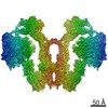 4805C 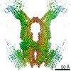 4806C 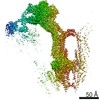 4807C  4808C  4810C 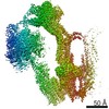 4811C  4812C  4813C 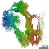 4814C 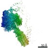 4815C 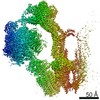 4816C  4817C  4818C 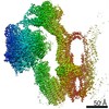 4819C  4820C  4821C  4822C  4823C  4824C  4825C  4826C  4827C  4828C  4829C  4830C  4831C  4832C  4833C  4834C  4835C  4836C  4837C  4838C  4839C  4840C  4841C  4842C  4843C  4844C  4845C 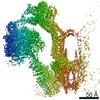 4846C 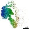 4847C 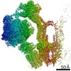 4848C  4849C 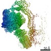 4850C 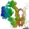 4851C  4852C  4853C  4854C  4855C  4856C 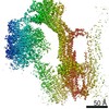 4857C 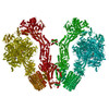 6rd4C  6rd5C  6rd6C 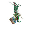 6rd7C  6rd9C  6rdaC 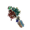 6rdbC  6rdcC  6rddC 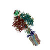 6rdeC 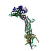 6rdfC  6rdgC 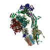 6rdhC  6rdiC 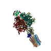 6rdjC 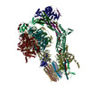 6rdkC  6rdlC 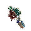 6rdmC  6rdnC 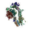 6rdoC  6rdpC 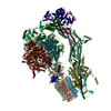 6rdqC  6rdrC  6rdsC  6rdtC  6rduC 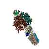 6rdvC  6rdwC 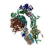 6rdxC  6rdyC 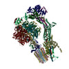 6rdzC  6re0C 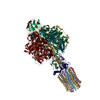 6re1C  6re2C 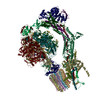 6re3C  6re4C 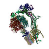 6re5C  6re6C  6re7C 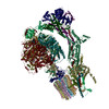 6re8C  6re9C  6reaC  6rebC 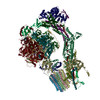 6recC  6redC 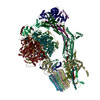 6reeC  6refC  6repC  6rerC  6resC  6retC 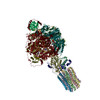 6reuC M: map data used to model this data C: citing same article ( |
|---|---|
| Similar structure data | |
| EM raw data | EMPIAR-10375 (Title: Rotary substates of mitochondrial ATP synthase reveal the basis of flexible F1-Fo coupling Data size: 43.8 TB Data #1: Unaligned frames, gain reference corrected [micrographs - multiframe]) |


-Links
 PDBj
PDBj

- Assembly
| Deposited unit | 
|
|---|---|
| 1 |
|
-Components
-Protein , 3 types, 3 molecules 019
| #1: Protein | Mass: 8731.866 Da / Num. of mol.: 1 / Source method: isolated from a natural source / Source: (natural) Polytomella sp. Pringsheim 198.80 (plant) / References: UniProt: A0A5H1ZR95*PLUS |
|---|---|
| #2: Protein | Mass: 68679.906 Da / Num. of mol.: 1 / Source method: isolated from a natural source / Source: (natural) Polytomella sp. Pringsheim 198.80 (plant) / References: UniProt: Q85JD5 |
| #7: Protein | Mass: 11001.712 Da / Num. of mol.: 1 / Source method: isolated from a natural source / Source: (natural) Polytomella sp. Pringsheim 198.80 (plant) / References: UniProt: A0A5H1ZR73*PLUS |
-Mitochondrial F1F0 ATP synthase associated ... , 2 types, 2 molecules 35
| #3: Protein | Mass: 34850.363 Da / Num. of mol.: 1 / Source method: isolated from a natural source / Source: (natural) Polytomella sp. Pringsheim 198.80 (plant) / References: UniProt: K0J903 |
|---|---|
| #4: Protein | Mass: 14004.376 Da / Num. of mol.: 1 / Source method: isolated from a natural source / Source: (natural) Polytomella sp. Pringsheim 198.80 (plant) / References: UniProt: A0A024FSR7 |
-Mitochondrial ATP synthase subunit ... , 4 types, 13 molecules 68ABCDEFGHIJM
| #5: Protein | Mass: 15904.290 Da / Num. of mol.: 1 / Source method: isolated from a natural source / Source: (natural) Polytomella sp. Pringsheim 198.80 (plant) / References: UniProt: D7P897 | ||
|---|---|---|---|
| #6: Protein | Mass: 9883.389 Da / Num. of mol.: 1 / Source method: isolated from a natural source / Source: (natural) Polytomella sp. Pringsheim 198.80 (plant) / References: UniProt: D8V7I7 | ||
| #8: Protein | Mass: 12664.013 Da / Num. of mol.: 10 / Source method: isolated from a natural source / Source: (natural) Polytomella sp. Pringsheim 198.80 (plant) / References: UniProt: D7P7X5#9: Protein | | Mass: 34802.344 Da / Num. of mol.: 1 / Source method: isolated from a natural source / Source: (natural) Polytomella sp. Pringsheim 198.80 (plant) / References: UniProt: H8PGG3 |
-Non-polymers , 2 types, 19 molecules 

| #10: Chemical | ChemComp-ZN /  Mass: 65.409 Da / Num. of mol.: 1 / Source method: obtained synthetically / Formula: Zn Mass: 65.409 Da / Num. of mol.: 1 / Source method: obtained synthetically / Formula: Zn |
|---|---|
| #11: Water | ChemComp-HOH / Mass: 18.015 Da / Num. of mol.: 18 / Source method: isolated from a natural source / Formula: H2O |
-Details
| Has protein modification | Y |
|---|
-Experimental details
-Experiment
| Experiment | Method: ELECTRON MICROSCOPY |
|---|---|
| EM experiment | Aggregation state: PARTICLE / 3D reconstruction method: single particle reconstruction |
- Sample preparation
| Component | Name: Polytomella F-ATP synthase subunits ASA1, ASA3, ASA5, ASA6, ASA8, ASA9, ASA10, c-ring and a-subunit. Type: COMPLEX / Entity ID: #1-#9 / Source: NATURAL |
|---|---|
| Source (natural) | Organism: Polytomella sp. Pringsheim 198.80 (plant) |
| Buffer solution | pH: 7.8 |
| Specimen | Conc.: 3.5 mg/ml / Embedding applied: NO / Shadowing applied: NO / Staining applied: NO / Vitrification applied: YES |
| Vitrification | Instrument: FEI VITROBOT MARK IV / Cryogen name: ETHANE / Humidity: 100 % / Chamber temperature: 277 K |
- Electron microscopy imaging
| Experimental equipment |  Model: Titan Krios / Image courtesy: FEI Company |
|---|---|
| Microscopy | Model: FEI TITAN KRIOS |
| Electron gun | Electron source:  FIELD EMISSION GUN / Accelerating voltage: 300 kV / Illumination mode: FLOOD BEAM FIELD EMISSION GUN / Accelerating voltage: 300 kV / Illumination mode: FLOOD BEAM |
| Electron lens | Mode: BRIGHT FIELD / Nominal magnification: 75000 X / Nominal defocus max: -5000 nm / Nominal defocus min: -400 nm / Cs: 2.7 mm / C2 aperture diameter: 70 µm / Alignment procedure: BASIC |
| Specimen holder | Cryogen: NITROGEN / Specimen holder model: FEI TITAN KRIOS AUTOGRID HOLDER |
| Image recording | Electron dose: 35 e/Å2 / Detector mode: COUNTING / Film or detector model: FEI FALCON III (4k x 4k) |
| Image scans | Width: 4096 / Height: 4096 |
- Processing
| EM software |
| ||||||||||||||||||||||||||||
|---|---|---|---|---|---|---|---|---|---|---|---|---|---|---|---|---|---|---|---|---|---|---|---|---|---|---|---|---|---|
| CTF correction | Type: PHASE FLIPPING AND AMPLITUDE CORRECTION | ||||||||||||||||||||||||||||
| Particle selection | Num. of particles selected: 735197 | ||||||||||||||||||||||||||||
| 3D reconstruction | Resolution: 3.08 Å / Resolution method: FSC 0.143 CUT-OFF / Num. of particles: 136422 / Symmetry type: POINT | ||||||||||||||||||||||||||||
| Atomic model building | Space: REAL |
