 Movie
Movie Controller


+ Open data

- Basic information
| Entry | Database: PDB / ID: 6r65 | |||||||||
|---|---|---|---|---|---|---|---|---|---|---|
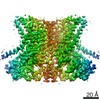
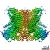
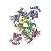
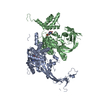
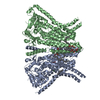
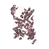
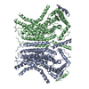
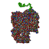
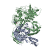
| Title | Crystal Structure of human TMEM16K / Anoctamin 10 (Form 2) | |||||||||
 Components Components | Anoctamin-10 | |||||||||
 Keywords Keywords | LIPID TRANSPORT / MEMBRANE PROTEIN / CALCIUM ACTIVATION / TRANSPORT PROTEIN / Structural Genomics / Structural Genomics Consortium / SGC | |||||||||
| Function / homology |  Function and homology information Function and homology informationintracellularly calcium-gated chloride channel activity / calcium-activated cation channel activity / chloride channel activity / chloride transmembrane transport / Stimuli-sensing channels / monoatomic ion transmembrane transport / Induction of Cell-Cell Fusion / cilium / membrane / plasma membrane Similarity search - Function | |||||||||
| Biological species |  Homo sapiens (human) Homo sapiens (human) | |||||||||
| Method |  X-RAY DIFFRACTION / SYNCHROTRON / MOLECULAR REPLACEMENT / Resolution: 3.5 Å X-RAY DIFFRACTION / SYNCHROTRON / MOLECULAR REPLACEMENT / Resolution: 3.5 Å | |||||||||
 Authors Authors | Bushell, S.R. / Pike, A.C.W. / Chu, A. / Tessitore, A. / Rotty, B. / Mukhopadhyay, S. / Kupinska, K. / Shrestha, L. / Borkowska, O. / Chalk, R. ...Bushell, S.R. / Pike, A.C.W. / Chu, A. / Tessitore, A. / Rotty, B. / Mukhopadhyay, S. / Kupinska, K. / Shrestha, L. / Borkowska, O. / Chalk, R. / Burgess-Brown, N.A. / Love, J. / Edwards, A.M. / Arrowsmith, C.H. / Bountra, C. / Carpenter, E.P. / Structural Genomics Consortium (SGC) | |||||||||
| Funding support |  United Kingdom, 2items United Kingdom, 2items
| |||||||||
 Citation Citation | Journal: Nat Commun / Year: 2019 Title: The structural basis of lipid scrambling and inactivation in the endoplasmic reticulum scramblase TMEM16K. Authors: Simon R Bushell / Ashley C W Pike / Maria E Falzone / Nils J G Rorsman / Chau M Ta / Robin A Corey / Thomas D Newport / John C Christianson / Lara F Scofano / Chitra A Shintre / Annamaria ...Authors: Simon R Bushell / Ashley C W Pike / Maria E Falzone / Nils J G Rorsman / Chau M Ta / Robin A Corey / Thomas D Newport / John C Christianson / Lara F Scofano / Chitra A Shintre / Annamaria Tessitore / Amy Chu / Qinrui Wang / Leela Shrestha / Shubhashish M M Mukhopadhyay / James D Love / Nicola A Burgess-Brown / Rebecca Sitsapesan / Phillip J Stansfeld / Juha T Huiskonen / Paolo Tammaro / Alessio Accardi / Elisabeth P Carpenter /   Abstract: Membranes in cells have defined distributions of lipids in each leaflet, controlled by lipid scramblases and flip/floppases. However, for some intracellular membranes such as the endoplasmic ...Membranes in cells have defined distributions of lipids in each leaflet, controlled by lipid scramblases and flip/floppases. However, for some intracellular membranes such as the endoplasmic reticulum (ER) the scramblases have not been identified. Members of the TMEM16 family have either lipid scramblase or chloride channel activity. Although TMEM16K is widely distributed and associated with the neurological disorder autosomal recessive spinocerebellar ataxia type 10 (SCAR10), its location in cells, function and structure are largely uncharacterised. Here we show that TMEM16K is an ER-resident lipid scramblase with a requirement for short chain lipids and calcium for robust activity. Crystal structures of TMEM16K show a scramblase fold, with an open lipid transporting groove. Additional cryo-EM structures reveal extensive conformational changes from the cytoplasmic to the ER side of the membrane, giving a state with a closed lipid permeation pathway. Molecular dynamics simulations showed that the open-groove conformation is necessary for scramblase activity. | |||||||||
| History |
|
- Structure visualization
| Structure viewer | Molecule: MolmilJmol/JSmol |
|---|
- Downloads & links
-Download
| PDBx/mmCIF format | 6r65.cif.gz | 494.1 KB | Display | PDBx/mmCIF format |
|---|---|---|---|---|
| PDB format | pdb6r65.ent.gz | 407.7 KB | Display | PDB format |
| PDBx/mmJSON format | 6r65.json.gz | Tree view | PDBx/mmJSON format | |
| Others |  Other downloads Other downloads |
-Validation report
| Arichive directory | https://data.pdbj.org/pub/pdb/validation_reports/r6/6r65ftp://data.pdbj.org/pub/pdb/validation_reports/r6/6r65 | HTTPS FTP |
|---|
-Related structure data
| Related structure data | 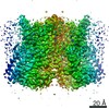 4746C 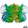 4747C 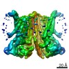 4748C  5oc9SC  6r7xC  6r7yC 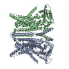 6r7zC S: Starting model for refinement C: citing same article ( |
|---|---|
| Similar structure data |

-Links
 PDBj
PDBj- Assembly
| Deposited unit | 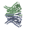
| ||||||||
|---|---|---|---|---|---|---|---|---|---|
| 1 |
| ||||||||
| 2 |
| ||||||||
| Unit cell |
|
-Components
| #1: Protein | Mass: 77276.016 Da / Num. of mol.: 2 Source method: isolated from a genetically manipulated source Source: (gene. exp.) Homo sapiens (human) / Gene: ANO10, TMEM16K / Plasmid: PFB-CT10HF-LIC / Production host:   Spodoptera frugiperda (fall armyworm) / References: UniProt: Q9NW15 Spodoptera frugiperda (fall armyworm) / References: UniProt: Q9NW15#2: Chemical | ChemComp-CA /   Mass: 40.078 Da / Num. of mol.: 6 / Source method: obtained synthetically / Formula: Ca Mass: 40.078 Da / Num. of mol.: 6 / Source method: obtained synthetically / Formula: Ca |
|---|
-Experimental details
-Experiment
| Experiment | Method: X-RAY DIFFRACTION / Number of used crystals: 1 |
|---|
- Sample preparation
| Crystal | Density Matthews: 4.34 Å3/Da / Density % sol: 71.66 % |
|---|---|
| Crystal grow | Temperature: 293 K / Method: vapor diffusion, sitting drop / pH: 7 Details: 0.1M calcium acetate 0.1M HEPES pH 7.0 22% PEG400 1CMC C12E9 |
-Data collection
| Diffraction | Mean temperature: 100 K / Serial crystal experiment: N | ||||||||||||||||||||||||||||||
|---|---|---|---|---|---|---|---|---|---|---|---|---|---|---|---|---|---|---|---|---|---|---|---|---|---|---|---|---|---|---|---|
| Diffraction source | Source: SYNCHROTRON / Site: Diamond / Beamline: I24 / Wavelength: 0.9686 Å | ||||||||||||||||||||||||||||||
| Detector | Type: DECTRIS PILATUS3 S 6M / Detector: PIXEL / Date: Nov 29, 2015 | ||||||||||||||||||||||||||||||
| Radiation | Protocol: SINGLE WAVELENGTH / Monochromatic (M) / Laue (L): M / Scattering type: x-ray | ||||||||||||||||||||||||||||||
| Radiation wavelength | Wavelength: 0.9686 Å / Relative weight: 1 | ||||||||||||||||||||||||||||||
| Reflection | Entry-ID: 6R65
| ||||||||||||||||||||||||||||||
| Reflection shell |
|
- Processing
| Software |
| ||||||||||||||||||||||||||||||||||||||||||||||||||||||||||||||||||||||||||||||||||||||||||||||||||||||||||||
|---|---|---|---|---|---|---|---|---|---|---|---|---|---|---|---|---|---|---|---|---|---|---|---|---|---|---|---|---|---|---|---|---|---|---|---|---|---|---|---|---|---|---|---|---|---|---|---|---|---|---|---|---|---|---|---|---|---|---|---|---|---|---|---|---|---|---|---|---|---|---|---|---|---|---|---|---|---|---|---|---|---|---|---|---|---|---|---|---|---|---|---|---|---|---|---|---|---|---|---|---|---|---|---|---|---|---|---|---|---|
| Refinement | Method to determine structure: MOLECULAR REPLACEMENT Starting model: 5OC9 Resolution: 3.5→39.69 Å / Cor.coef. Fo:Fc: 0.817 / Cor.coef. Fo:Fc free: 0.794 / Cross valid method: THROUGHOUT / σ(F): 0 / SU Rfree Blow DPI: 0.605 Details: Structure refined using STARANISO anistropically truncated data. Reference model (LSSR) restraints to PDB:5OC9 were also used
| ||||||||||||||||||||||||||||||||||||||||||||||||||||||||||||||||||||||||||||||||||||||||||||||||||||||||||||
| Displacement parameters | Biso max: 229.57 Å2 / Biso mean: 92.15 Å2 / Biso min: 57.28 Å2
| ||||||||||||||||||||||||||||||||||||||||||||||||||||||||||||||||||||||||||||||||||||||||||||||||||||||||||||
| Refine analyze | Luzzati coordinate error obs: 0.59 Å | ||||||||||||||||||||||||||||||||||||||||||||||||||||||||||||||||||||||||||||||||||||||||||||||||||||||||||||
| Refinement step | Cycle: final / Resolution: 3.5→39.69 Å
| ||||||||||||||||||||||||||||||||||||||||||||||||||||||||||||||||||||||||||||||||||||||||||||||||||||||||||||
| Refine LS restraints |
| ||||||||||||||||||||||||||||||||||||||||||||||||||||||||||||||||||||||||||||||||||||||||||||||||||||||||||||
| LS refinement shell | Resolution: 3.5→3.65 Å / Rfactor Rfree error: 0 / Total num. of bins used: 12
| ||||||||||||||||||||||||||||||||||||||||||||||||||||||||||||||||||||||||||||||||||||||||||||||||||||||||||||
| Refinement TLS params. | Method: refined / Refine-ID: X-RAY DIFFRACTION
| ||||||||||||||||||||||||||||||||||||||||||||||||||||||||||||||||||||||||||||||||||||||||||||||||||||||||||||
| Refinement TLS group |
|
