 Movie
Movie Controller

[English] 日本語
 Yorodumi
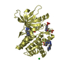
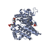
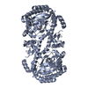
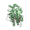
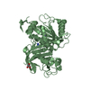
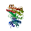
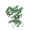
- PDB-6r61: Major aspartyl peptidase 1 from C. neoformans in complex with Inh... -
+ Open data

- Basic information
| Entry | Database: PDB / ID: 6r61 | ||||||
|---|---|---|---|---|---|---|---|
| Title | Major aspartyl peptidase 1 from C. neoformans in complex with Inhibitor LP258 | ||||||
 Components Components | Endopeptidase | ||||||
 Keywords Keywords | HYDROLASE / aspartyl protease / secreted / Cryptococcus neoformans / inhibitor complex | ||||||
| Function / homology |  Function and homology information Function and homology information | ||||||
| Biological species |  Cryptococcus neoformans (fungus) Cryptococcus neoformans (fungus) | ||||||
| Method |  X-RAY DIFFRACTION / FOURIER SYNTHESIS / Resolution: 1.81 Å X-RAY DIFFRACTION / FOURIER SYNTHESIS / Resolution: 1.81 Å | ||||||
 Authors Authors | Krystufek, R. / Sacha, P. / Brynda, J. / Konvalinka, J. | ||||||
| Funding support |  Czech Republic, 1items Czech Republic, 1items
| ||||||
 Citation Citation | Journal: To Be Published Title: Major aspartyl peptidase 1 from C. neoformans in complex with Inhibitor LP258 Authors: Krystufek, R. / Sacha, P. / Brynda, J. / Konvalinka, J. | ||||||
| History |
|
- Structure visualization
| Structure viewer | Molecule: MolmilJmol/JSmol |
|---|
- Downloads & links
-Download
| PDBx/mmCIF format | 6r61.cif.gz | 91.6 KB | Display | PDBx/mmCIF format |
|---|---|---|---|---|
| PDB format | pdb6r61.ent.gz | 65.1 KB | Display | PDB format |
| PDBx/mmJSON format | 6r61.json.gz | Tree view | PDBx/mmJSON format | |
| Others |  Other downloads Other downloads |
-Validation report
| Summary document | 6r61_validation.pdf.gz | 931.7 KB | Display | wwPDB validaton report |
|---|---|---|---|---|
| Full document | 6r61_full_validation.pdf.gz | 930 KB | Display | |
| Data in XML | 6r61_validation.xml.gz | 17.3 KB | Display | |
| Arichive directory | https://data.pdbj.org/pub/pdb/validation_reports/r6/6r61ftp://data.pdbj.org/pub/pdb/validation_reports/r6/6r61 | HTTPS FTP |
-Related structure data
| Related structure data | 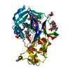 6r5hS S: Starting model for refinement |
|---|---|
| Similar structure data |


-Links
 PDBj
PDBj- Assembly
| Deposited unit | 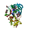
| |||||||||
|---|---|---|---|---|---|---|---|---|---|---|
| 1 |
| |||||||||
| Unit cell |
| |||||||||
| Components on special symmetry positions |
|
-Components
-Protein / Sugars , 2 types, 2 molecules A
| #1: Protein | Mass: 36811.281 Da / Num. of mol.: 1 Source method: isolated from a genetically manipulated source Details: MayI(17-434)-Avitag / Source: (gene. exp.) Cryptococcus neoformans (fungus) / Gene: CNAG_05872 / Plasmid: pMT_BIP_MayI(17-434)Avitag / Production host:  |
|---|---|
| #2: Polysaccharide | 2-acetamido-2-deoxy-beta-D-glucopyranose-(1-4)-2-acetamido-2-deoxy-beta-D-glucopyranose Source method: isolated from a genetically manipulated source |
-Non-polymers , 7 types, 202 molecules 


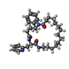

| #3: Chemical |  Mass: 150.173 Da / Num. of mol.: 3 / Source method: obtained synthetically / Formula: C6H14O4 Mass: 150.173 Da / Num. of mol.: 3 / Source method: obtained synthetically / Formula: C6H14O4#4: Chemical | ChemComp-PEG /  Mass: 106.120 Da / Num. of mol.: 4 / Source method: obtained synthetically / Formula: C4H10O3 Mass: 106.120 Da / Num. of mol.: 4 / Source method: obtained synthetically / Formula: C4H10O3#5: Chemical | ChemComp-SO4 / |  Mass: 96.063 Da / Num. of mol.: 1 / Source method: obtained synthetically / Formula: SO4 Mass: 96.063 Da / Num. of mol.: 1 / Source method: obtained synthetically / Formula: SO4#6: Chemical | ChemComp-CL / |  Mass: 35.453 Da / Num. of mol.: 1 / Source method: obtained synthetically / Formula: Cl Mass: 35.453 Da / Num. of mol.: 1 / Source method: obtained synthetically / Formula: Cl#7: Chemical | ChemComp-NA / |  Mass: 22.990 Da / Num. of mol.: 1 / Source method: obtained synthetically / Formula: Na Mass: 22.990 Da / Num. of mol.: 1 / Source method: obtained synthetically / Formula: Na#8: Chemical | ChemComp-JT8 / ( |  Mass: 564.759 Da / Num. of mol.: 1 / Source method: obtained synthetically / Formula: C33H48N4O4 Mass: 564.759 Da / Num. of mol.: 1 / Source method: obtained synthetically / Formula: C33H48N4O4#9: Water | ChemComp-HOH / | Mass: 18.015 Da / Num. of mol.: 191 / Source method: isolated from a natural source / Formula: H2O |
|---|
-Experimental details
-Experiment
| Experiment | Method: X-RAY DIFFRACTION / Number of used crystals: 1 |
|---|
- Sample preparation
| Crystal | Density Matthews: 3.41 Å3/Da / Density % sol: 63.96 % |
|---|---|
| Crystal grow | Temperature: 298 K / Method: vapor diffusion, hanging drop Details: peptidase (85 mg/ml) in 50 mM sodium acetate, pH=5.0, 100mM NaCl in equal volume with reservoir solution: 100mM lithium sulphate, 45% (v/v) PEG-400, 100 mM sodium acetate pH= 4.5 PH range: 4.5 - 5.0 |
-Data collection
| Diffraction | Mean temperature: 100 K / Serial crystal experiment: N | ||||||||||||||||||||||||||||||||||||||||||||||||||||||||||||||||||||||||||||||||||||||||||||||||||||||||||||||||||||||||||||||||||||||||||||||||||||||||||||||||||||||||
|---|---|---|---|---|---|---|---|---|---|---|---|---|---|---|---|---|---|---|---|---|---|---|---|---|---|---|---|---|---|---|---|---|---|---|---|---|---|---|---|---|---|---|---|---|---|---|---|---|---|---|---|---|---|---|---|---|---|---|---|---|---|---|---|---|---|---|---|---|---|---|---|---|---|---|---|---|---|---|---|---|---|---|---|---|---|---|---|---|---|---|---|---|---|---|---|---|---|---|---|---|---|---|---|---|---|---|---|---|---|---|---|---|---|---|---|---|---|---|---|---|---|---|---|---|---|---|---|---|---|---|---|---|---|---|---|---|---|---|---|---|---|---|---|---|---|---|---|---|---|---|---|---|---|---|---|---|---|---|---|---|---|---|---|---|---|---|---|---|---|
| Diffraction source | Source: ROTATING ANODE / Type: RIGAKU MICROMAX-007 HF / Wavelength: 0.9184 Å | ||||||||||||||||||||||||||||||||||||||||||||||||||||||||||||||||||||||||||||||||||||||||||||||||||||||||||||||||||||||||||||||||||||||||||||||||||||||||||||||||||||||||
| Detector | Type: DECTRIS PILATUS 300K / Detector: PIXEL / Date: Sep 11, 2018 | ||||||||||||||||||||||||||||||||||||||||||||||||||||||||||||||||||||||||||||||||||||||||||||||||||||||||||||||||||||||||||||||||||||||||||||||||||||||||||||||||||||||||
| Radiation | Monochromator: Confocal Max-Flux optics Rigaku / Protocol: SINGLE WAVELENGTH / Monochromatic (M) / Laue (L): M / Scattering type: x-ray | ||||||||||||||||||||||||||||||||||||||||||||||||||||||||||||||||||||||||||||||||||||||||||||||||||||||||||||||||||||||||||||||||||||||||||||||||||||||||||||||||||||||||
| Radiation wavelength | Wavelength: 0.9184 Å / Relative weight: 1 | ||||||||||||||||||||||||||||||||||||||||||||||||||||||||||||||||||||||||||||||||||||||||||||||||||||||||||||||||||||||||||||||||||||||||||||||||||||||||||||||||||||||||
| Reflection | Resolution: 1.81→73.78 Å / Num. obs: 44101 / % possible obs: 95.8 % / Redundancy: 3.764 % / Biso Wilson estimate: 24.145 Å2 / CC1/2: 0.991 / Rmerge(I) obs: 0.175 / Rrim(I) all: 0.202 / Χ2: 0.892 / Net I/σ(I): 6.66 | ||||||||||||||||||||||||||||||||||||||||||||||||||||||||||||||||||||||||||||||||||||||||||||||||||||||||||||||||||||||||||||||||||||||||||||||||||||||||||||||||||||||||
| Reflection shell | Diffraction-ID: 1
|
- Processing
| Software |
| ||||||||||||||||||||||||||||||||||||||||||||||||||||||||||||
|---|---|---|---|---|---|---|---|---|---|---|---|---|---|---|---|---|---|---|---|---|---|---|---|---|---|---|---|---|---|---|---|---|---|---|---|---|---|---|---|---|---|---|---|---|---|---|---|---|---|---|---|---|---|---|---|---|---|---|---|---|---|
| Refinement | Method to determine structure: FOURIER SYNTHESIS Starting model: 6R5H Resolution: 1.81→73.78 Å / Cor.coef. Fo:Fc: 0.956 / Cor.coef. Fo:Fc free: 0.951 / SU B: 2.849 / SU ML: 0.081 / SU R Cruickshank DPI: 0.1073 / Cross valid method: THROUGHOUT / σ(F): 0 / ESU R: 0.107 / ESU R Free: 0.099 Details: HYDROGENS HAVE BEEN ADDED IN THE RIDING POSITIONS U VALUES : REFINED INDIVIDUALLY
| ||||||||||||||||||||||||||||||||||||||||||||||||||||||||||||
| Solvent computation | Ion probe radii: 0.8 Å / Shrinkage radii: 0.8 Å / VDW probe radii: 1.2 Å | ||||||||||||||||||||||||||||||||||||||||||||||||||||||||||||
| Displacement parameters | Biso max: 115.89 Å2 / Biso mean: 26.587 Å2 / Biso min: 8.6 Å2
| ||||||||||||||||||||||||||||||||||||||||||||||||||||||||||||
| Refinement step | Cycle: final / Resolution: 1.81→73.78 Å
| ||||||||||||||||||||||||||||||||||||||||||||||||||||||||||||
| Refine LS restraints |
| ||||||||||||||||||||||||||||||||||||||||||||||||||||||||||||
| LS refinement shell | Resolution: 1.81→1.857 Å / Rfactor Rfree error: 0 / Total num. of bins used: 20
|
