 Movie
Movie Controller


+ Open data

- Basic information
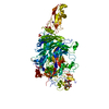
| Entry | Database: PDB / ID: 6r3t | |||||||||
|---|---|---|---|---|---|---|---|---|---|---|
| Title | Structure of P110 from Mycoplasma Genitalium at 2.7A | |||||||||
 Components Components | Mgp-operon protein 3 | |||||||||
 Keywords Keywords | SUGAR BINDING PROTEIN / Adhesin | |||||||||
| Function / homology | MgpC adhesin / Mgp-operon protein 3, C-terminal domain / MgpC adhesin / MGP3 C-terminal domain / cell adhesion / plasma membrane / : / PHOSPHATE ION / Adhesin P110 Function and homology information Function and homology information | |||||||||
| Biological species |  Mycoplasma genitalium G37 (bacteria) Mycoplasma genitalium G37 (bacteria) | |||||||||
| Method |  X-RAY DIFFRACTION / SYNCHROTRON / SAD / Resolution: 2.73 Å X-RAY DIFFRACTION / SYNCHROTRON / SAD / Resolution: 2.73 Å | |||||||||
 Authors Authors | Aparicio, D. / Fita, I. | |||||||||
 Citation Citation | Journal: Nat Commun / Year: 2018 Title: Mycoplasma genitalium adhesin P110 binds sialic-acid human receptors. Authors: Aparicio, D. / Torres-Puig, S. / Ratera, M. / Querol, E. / Pinol, J. / Pich, O.Q. / Fita, I. | |||||||||
| History |
|
- Structure visualization
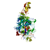
| Structure viewer | Molecule: MolmilJmol/JSmol |
|---|
- Downloads & links
-Download
| PDBx/mmCIF format | 6r3t.cif.gz | 363.8 KB | Display | PDBx/mmCIF format |
|---|---|---|---|---|
| PDB format | pdb6r3t.ent.gz | 298.7 KB | Display | PDB format |
| PDBx/mmJSON format | 6r3t.json.gz | Tree view | PDBx/mmJSON format | |
| Others |  Other downloads Other downloads |
-Validation report
| Arichive directory | https://data.pdbj.org/pub/pdb/validation_reports/r3/6r3tftp://data.pdbj.org/pub/pdb/validation_reports/r3/6r3t | HTTPS FTP |
|---|
-Related structure data


-Links
 PDBj
PDBj- Assembly
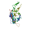
| Deposited unit | 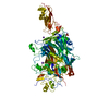
| ||||||||
|---|---|---|---|---|---|---|---|---|---|
| 1 |
| ||||||||
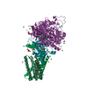
| Unit cell |
|
-Components
| #1: Protein | Mass: 99555.203 Da / Num. of mol.: 1 Source method: isolated from a genetically manipulated source Source: (gene. exp.) Mycoplasma genitalium G37 (bacteria) / Gene: MG192 / Production host: | ||
|---|---|---|---|
| #2: Chemical | ChemComp-PO4 /   Mass: 94.971 Da / Num. of mol.: 1 / Source method: obtained synthetically / Formula: PO4 Mass: 94.971 Da / Num. of mol.: 1 / Source method: obtained synthetically / Formula: PO4 | ||
| #3: Chemical |   Mass: 39.098 Da / Num. of mol.: 2 / Source method: obtained synthetically / Formula: K Mass: 39.098 Da / Num. of mol.: 2 / Source method: obtained synthetically / Formula: K#4: Water | ChemComp-HOH / |  Mass: 18.015 Da / Num. of mol.: 63 / Source method: isolated from a natural source / Formula: H2O Mass: 18.015 Da / Num. of mol.: 63 / Source method: isolated from a natural source / Formula: H2O |
-Experimental details
-Experiment
| Experiment | Method: X-RAY DIFFRACTION / Number of used crystals: 1 |
|---|
- Sample preparation
| Crystal | Density Matthews: 3.6 Å3/Da / Density % sol: 65.88 % |
|---|---|
| Crystal grow | Temperature: 293 K / Method: vapor diffusion, hanging drop Details: 11% PEG 8000 0.1M Imidazole pH 8 0.2M Calcium acetate |
-Data collection
| Diffraction | Mean temperature: 293 K / Serial crystal experiment: N |
|---|---|
| Diffraction source | Source: SYNCHROTRON / Site: ALBA  / Beamline: XALOC / Wavelength: 1.12712 Å / Beamline: XALOC / Wavelength: 1.12712 Å |
| Detector | Type: DECTRIS PILATUS 6M / Detector: PIXEL / Date: Mar 10, 2016 |
| Radiation | Protocol: SINGLE WAVELENGTH / Monochromatic (M) / Laue (L): M / Scattering type: x-ray |
| Radiation wavelength | Wavelength: 1.12712 Å / Relative weight: 1 |
| Reflection | Resolution: 2.7→62 Å / Num. obs: 39641 / % possible obs: 98.9 % / Redundancy: 4.6 % / Rmerge(I) obs: 0.047 / Rpim(I) all: 0.052 / Net I/σ(I): 19.1 |
| Reflection shell | Resolution: 2.7→2.77 Å / Redundancy: 4.8 % / Num. unique obs: 39641 |
- Processing
| Software |
| ||||||||||||||||||||||||||||||||||||||||||||||||||||||||||||||||||||||||||||||||||||||||||||||||||||||||||||||||||||||||||||||||||||||||||||||||||||||||||||||||||||||||||||||||||||||
|---|---|---|---|---|---|---|---|---|---|---|---|---|---|---|---|---|---|---|---|---|---|---|---|---|---|---|---|---|---|---|---|---|---|---|---|---|---|---|---|---|---|---|---|---|---|---|---|---|---|---|---|---|---|---|---|---|---|---|---|---|---|---|---|---|---|---|---|---|---|---|---|---|---|---|---|---|---|---|---|---|---|---|---|---|---|---|---|---|---|---|---|---|---|---|---|---|---|---|---|---|---|---|---|---|---|---|---|---|---|---|---|---|---|---|---|---|---|---|---|---|---|---|---|---|---|---|---|---|---|---|---|---|---|---|---|---|---|---|---|---|---|---|---|---|---|---|---|---|---|---|---|---|---|---|---|---|---|---|---|---|---|---|---|---|---|---|---|---|---|---|---|---|---|---|---|---|---|---|---|---|---|---|---|
| Refinement | Method to determine structure: SAD / Resolution: 2.73→54.86 Å / Cor.coef. Fo:Fc: 0.963 / Cor.coef. Fo:Fc free: 0.941 / SU B: 27.881 / SU ML: 0.254 / Cross valid method: THROUGHOUT / ESU R: 0.433 / ESU R Free: 0.273 / Details: HYDROGENS HAVE BEEN ADDED IN THE RIDING POSITIONS
| ||||||||||||||||||||||||||||||||||||||||||||||||||||||||||||||||||||||||||||||||||||||||||||||||||||||||||||||||||||||||||||||||||||||||||||||||||||||||||||||||||||||||||||||||||||||
| Solvent computation | Ion probe radii: 0.8 Å / Shrinkage radii: 0.8 Å / VDW probe radii: 1.2 Å | ||||||||||||||||||||||||||||||||||||||||||||||||||||||||||||||||||||||||||||||||||||||||||||||||||||||||||||||||||||||||||||||||||||||||||||||||||||||||||||||||||||||||||||||||||||||
| Displacement parameters | Biso mean: 89.398 Å2
| ||||||||||||||||||||||||||||||||||||||||||||||||||||||||||||||||||||||||||||||||||||||||||||||||||||||||||||||||||||||||||||||||||||||||||||||||||||||||||||||||||||||||||||||||||||||
| Refinement step | Cycle: 1 / Resolution: 2.73→54.86 Å
| ||||||||||||||||||||||||||||||||||||||||||||||||||||||||||||||||||||||||||||||||||||||||||||||||||||||||||||||||||||||||||||||||||||||||||||||||||||||||||||||||||||||||||||||||||||||
| Refine LS restraints |
|