 Movie
Movie Controller

[English] 日本語
 Yorodumi
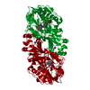
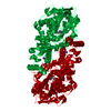
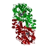
- PDB-6r3i: Aspergillus niger ferulic acid decarboxylase (Fdc) E282Q variant ... -
+ Open data

- Basic information
| Entry | Database: PDB / ID: 6r3i | |||||||||
|---|---|---|---|---|---|---|---|---|---|---|
| Title | Aspergillus niger ferulic acid decarboxylase (Fdc) E282Q variant in complex with the covalent adduct formed between prFMN cofactor and pentafluorocinnamic acid (Int2) | |||||||||
 Components Components | Ferulic acid decarboxylase 1 | |||||||||
 Keywords Keywords | LYASE / (de)carboxylase / UbiD / prFMN binding | |||||||||
| Function / homology |  Function and homology information Function and homology informationstyrene metabolic process / aromatic amino acid catabolic process / phenacrylate decarboxylase / ferulate metabolic process / cinnamic acid catabolic process / carboxy-lyase activity / manganese ion binding / identical protein binding / cytoplasm Similarity search - Function | |||||||||
| Biological species |  | |||||||||
| Method |  X-RAY DIFFRACTION / SYNCHROTRON / FOURIER SYNTHESIS / Resolution: 1.14 Å X-RAY DIFFRACTION / SYNCHROTRON / FOURIER SYNTHESIS / Resolution: 1.14 Å | |||||||||
 Authors Authors | Bailey, S.S. / Leys, D. | |||||||||
| Funding support |  United Kingdom, 2items United Kingdom, 2items
| |||||||||
 Citation Citation | Journal: To be published Title: Atomic description of an enzyme reaction dependent on reversible 1,3-dipolar cycloaddition Authors: Bailey, S.S. / Payne, K.A.P. / Saaret, A. / Marshall, S.A. / Gostimskaya, I. / Kosov, I. / Fisher, K. / Hay, S. / Leys, D. | |||||||||
| History |
|
- Structure visualization
| Structure viewer | Molecule: MolmilJmol/JSmol |
|---|
- Downloads & links
-Download
| PDBx/mmCIF format | 6r3i.cif.gz | 238.5 KB | Display | PDBx/mmCIF format |
|---|---|---|---|---|
| PDB format | pdb6r3i.ent.gz | 188.4 KB | Display | PDB format |
| PDBx/mmJSON format | 6r3i.json.gz | Tree view | PDBx/mmJSON format | |
| Others |  Other downloads Other downloads |
-Validation report
| Arichive directory | https://data.pdbj.org/pub/pdb/validation_reports/r3/6r3iftp://data.pdbj.org/pub/pdb/validation_reports/r3/6r3i | HTTPS FTP |
|---|
-Related structure data
| Related structure data |  6r2pC 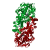 6r2rC  6r2tC  6r2zC  6r30C  6r32C  6r33C  6r34C  6r3fC  6r3gC  6r3jC  6r3lC  6r3nC  6r3oC  4za9S S: Starting model for refinement C: citing same article ( |
|---|---|
| Similar structure data |


-Links
 PDBj
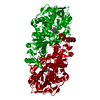
PDBj- Assembly
| Deposited unit | 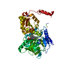
| ||||||||
|---|---|---|---|---|---|---|---|---|---|
| 1 | 
| ||||||||
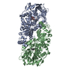
| Unit cell |
| ||||||||
| Components on special symmetry positions |
|
-Components
| #1: Protein | Mass: 56334.961 Da / Num. of mol.: 1 / Mutation: E282Q Source method: isolated from a genetically manipulated source Source: (gene. exp.)  | ||||
|---|---|---|---|---|---|
| #2: Chemical | ChemComp-MN /   Mass: 54.938 Da / Num. of mol.: 1 / Source method: obtained synthetically / Formula: Mn Mass: 54.938 Da / Num. of mol.: 1 / Source method: obtained synthetically / Formula: Mn | ||||
| #3: Chemical |   Mass: 39.098 Da / Num. of mol.: 2 / Source method: obtained synthetically / Formula: K Mass: 39.098 Da / Num. of mol.: 2 / Source method: obtained synthetically / Formula: K#4: Chemical | ChemComp-JRN / | 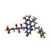  Mass: 720.578 Da / Num. of mol.: 1 / Source method: obtained synthetically / Formula: C30H34F5N4O9P / Feature type: SUBJECT OF INVESTIGATION Mass: 720.578 Da / Num. of mol.: 1 / Source method: obtained synthetically / Formula: C30H34F5N4O9P / Feature type: SUBJECT OF INVESTIGATION#5: Water | ChemComp-HOH / |  Mass: 18.015 Da / Num. of mol.: 446 / Source method: isolated from a natural source / Formula: H2O Mass: 18.015 Da / Num. of mol.: 446 / Source method: isolated from a natural source / Formula: H2O |
-Experimental details
-Experiment
| Experiment | Method: X-RAY DIFFRACTION / Number of used crystals: 1 |
|---|
- Sample preparation
| Crystal | Density Matthews: 2.39 Å3/Da / Density % sol: 48.62 % |
|---|---|
| Crystal grow | Temperature: 277 K / Method: vapor diffusion, sitting drop Details: 0.2 M POTASSIUM THIOCYANATE, BIS-TRIS PROPANE 6.5, 20 % W/V PEG 3350 |
-Data collection
| Diffraction | Mean temperature: 100 K / Serial crystal experiment: N |
|---|---|
| Diffraction source | Source: SYNCHROTRON / Site: Diamond / Beamline: I03 / Wavelength: 0.976 Å |
| Detector | Type: DECTRIS PILATUS 300K / Detector: PIXEL / Date: Aug 4, 2018 |
| Radiation | Protocol: SINGLE WAVELENGTH / Monochromatic (M) / Laue (L): M / Scattering type: x-ray |
| Radiation wavelength | Wavelength: 0.976 Å / Relative weight: 1 |
| Reflection | Resolution: 1.14→64.73 Å / Num. obs: 195884 / % possible obs: 99.56 % / Redundancy: 4.8 % / CC1/2: 0.997 / Net I/σ(I): 9.05 |
| Reflection shell | Resolution: 1.14→1.18 Å / Mean I/σ(I) obs: 2.09 / Num. unique obs: 19153 / CC1/2: 0.785 / % possible all: 98.3 |
- Processing
| Software |
| ||||||||||||||||||||||||||||||||||||||||||||||||||||||||||||||||||||||||||||||||||||||||||||||||||||||||||||||||||||||||||||||||||||||||||||||||||||||||||||||||||||||||||||||||||||||
|---|---|---|---|---|---|---|---|---|---|---|---|---|---|---|---|---|---|---|---|---|---|---|---|---|---|---|---|---|---|---|---|---|---|---|---|---|---|---|---|---|---|---|---|---|---|---|---|---|---|---|---|---|---|---|---|---|---|---|---|---|---|---|---|---|---|---|---|---|---|---|---|---|---|---|---|---|---|---|---|---|---|---|---|---|---|---|---|---|---|---|---|---|---|---|---|---|---|---|---|---|---|---|---|---|---|---|---|---|---|---|---|---|---|---|---|---|---|---|---|---|---|---|---|---|---|---|---|---|---|---|---|---|---|---|---|---|---|---|---|---|---|---|---|---|---|---|---|---|---|---|---|---|---|---|---|---|---|---|---|---|---|---|---|---|---|---|---|---|---|---|---|---|---|---|---|---|---|---|---|---|---|---|---|
| Refinement | Method to determine structure: FOURIER SYNTHESIS Starting model: 4ZA9 Resolution: 1.14→64.73 Å / Cor.coef. Fo:Fc: 0.98 / Cor.coef. Fo:Fc free: 0.974 / SU B: 0.885 / SU ML: 0.018 / Cross valid method: THROUGHOUT / ESU R: 0.026 / ESU R Free: 0.027 / Details: HYDROGENS HAVE BEEN ADDED IN THE RIDING POSITIONS
| ||||||||||||||||||||||||||||||||||||||||||||||||||||||||||||||||||||||||||||||||||||||||||||||||||||||||||||||||||||||||||||||||||||||||||||||||||||||||||||||||||||||||||||||||||||||
| Solvent computation | Ion probe radii: 0.8 Å / Shrinkage radii: 0.8 Å / VDW probe radii: 1.2 Å | ||||||||||||||||||||||||||||||||||||||||||||||||||||||||||||||||||||||||||||||||||||||||||||||||||||||||||||||||||||||||||||||||||||||||||||||||||||||||||||||||||||||||||||||||||||||
| Displacement parameters | Biso mean: 11.812 Å2
| ||||||||||||||||||||||||||||||||||||||||||||||||||||||||||||||||||||||||||||||||||||||||||||||||||||||||||||||||||||||||||||||||||||||||||||||||||||||||||||||||||||||||||||||||||||||
| Refinement step | Cycle: 1 / Resolution: 1.14→64.73 Å
| ||||||||||||||||||||||||||||||||||||||||||||||||||||||||||||||||||||||||||||||||||||||||||||||||||||||||||||||||||||||||||||||||||||||||||||||||||||||||||||||||||||||||||||||||||||||
| Refine LS restraints |
|