 Movie
Movie Controller

[English] 日本語
 Yorodumi
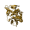
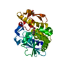
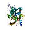
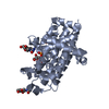
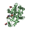
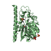
- PDB-6qw8: Crystal structure of CTX-M-15 complexed with relebactam (16 hour soak) -
+ Open data

- Basic information
| Entry | Database: PDB / ID: 6qw8 | ||||||
|---|---|---|---|---|---|---|---|
| Title | Crystal structure of CTX-M-15 complexed with relebactam (16 hour soak) | ||||||
 Components Components | Beta-lactamase | ||||||
 Keywords Keywords | ANTIMICROBIAL PROTEIN / inhibitor / relebactam / diazabicyclooctane / antibiotic resistance. | ||||||
| Function / homology |  Function and homology information Function and homology informationbeta-lactam antibiotic catabolic process / beta-lactamase activity / beta-lactamase / response to antibiotic / membrane Similarity search - Function | ||||||
| Biological species |  Klebsiella pneumoniae (bacteria) Klebsiella pneumoniae (bacteria) | ||||||
| Method |  X-RAY DIFFRACTION / SYNCHROTRON / MOLECULAR REPLACEMENT / Resolution: 1.1 Å X-RAY DIFFRACTION / SYNCHROTRON / MOLECULAR REPLACEMENT / Resolution: 1.1 Å | ||||||
 Authors Authors | Tooke, C.L. / Hinchliffe, P. / Spencer, J. | ||||||
| Funding support |  United Kingdom, 1items United Kingdom, 1items
| ||||||
 Citation Citation | Journal: Antimicrob.Agents Chemother. / Year: 2019 Title: Molecular Basis of Class A beta-Lactamase Inhibition by Relebactam. Authors: Tooke, C.L. / Hinchliffe, P. / Lang, P.A. / Mulholland, A.J. / Brem, J. / Schofield, C.J. / Spencer, J. | ||||||
| History |
|
- Structure visualization
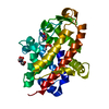
| Structure viewer | Molecule: MolmilJmol/JSmol |
|---|
- Downloads & links
-Download
| PDBx/mmCIF format | 6qw8.cif.gz | 179.4 KB | Display | PDBx/mmCIF format |
|---|---|---|---|---|
| PDB format | pdb6qw8.ent.gz | 142.9 KB | Display | PDB format |
| PDBx/mmJSON format | 6qw8.json.gz | Tree view | PDBx/mmJSON format | |
| Others |  Other downloads Other downloads |
-Validation report
| Summary document | 6qw8_validation.pdf.gz | 1 MB | Display | wwPDB validaton report |
|---|---|---|---|---|
| Full document | 6qw8_full_validation.pdf.gz | 1 MB | Display | |
| Data in XML | 6qw8_validation.xml.gz | 16 KB | Display | |
| Data in CIF | 6qw8_validation.cif.gz | 24.9 KB | Display | |
| Arichive directory | https://data.pdbj.org/pub/pdb/validation_reports/qw/6qw8ftp://data.pdbj.org/pub/pdb/validation_reports/qw/6qw8 | HTTPS FTP |
-Related structure data
| Related structure data |  6qw7C 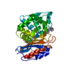 6qw9C 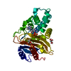 6qwaC 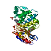 6qwbC 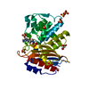 6qwcC 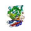 6qwdC  6qweC 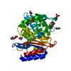 4hbtS S: Starting model for refinement C: citing same article ( |
|---|---|
| Similar structure data |

-Links
 PDBj
PDBj


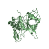
- Assembly
| Deposited unit | 
| ||||||||
|---|---|---|---|---|---|---|---|---|---|
| 1 |
| ||||||||
| Unit cell |
|
-Components
| #1: Protein | Mass: 28292.990 Da / Num. of mol.: 1 Source method: isolated from a genetically manipulated source Source: (gene. exp.) Klebsiella pneumoniae (bacteria) / Gene: blaCTX-M-15 / Production host: References: UniProt: G3G192, UniProt: W1EPV7*PLUS, beta-lactamase | ||||||
|---|---|---|---|---|---|---|---|
| #2: Chemical | ChemComp-MK7 / (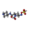  Mass: 350.391 Da / Num. of mol.: 1 / Source method: obtained synthetically / Formula: C12H22N4O6S / Feature type: SUBJECT OF INVESTIGATION Mass: 350.391 Da / Num. of mol.: 1 / Source method: obtained synthetically / Formula: C12H22N4O6S / Feature type: SUBJECT OF INVESTIGATION | ||||||
| #3: Chemical |   Mass: 96.063 Da / Num. of mol.: 3 / Source method: obtained synthetically / Formula: SO4 Mass: 96.063 Da / Num. of mol.: 3 / Source method: obtained synthetically / Formula: SO4#4: Chemical | ChemComp-GOL / |   Mass: 92.094 Da / Num. of mol.: 1 / Source method: obtained synthetically / Formula: C3H8O3 Mass: 92.094 Da / Num. of mol.: 1 / Source method: obtained synthetically / Formula: C3H8O3#5: Water | ChemComp-HOH / |  Mass: 18.015 Da / Num. of mol.: 368 / Source method: isolated from a natural source / Formula: H2O Mass: 18.015 Da / Num. of mol.: 368 / Source method: isolated from a natural source / Formula: H2OHas protein modification | Y | |
-Experimental details
-Experiment
| Experiment | Method: X-RAY DIFFRACTION / Number of used crystals: 1 |
|---|
- Sample preparation
| Crystal | Density Matthews: 2.11 Å3/Da / Density % sol: 41.83 % |
|---|---|
| Crystal grow | Temperature: 294.15 K / Method: vapor diffusion, sitting drop / Details: 2.0 M ammonium sulphate, 0.1 M Tris 8.0 |
-Data collection
| Diffraction | Mean temperature: 100 K / Serial crystal experiment: N |
|---|---|
| Diffraction source | Source: SYNCHROTRON / Site: Diamond / Beamline: I03 / Wavelength: 0.98 Å |
| Detector | Type: DECTRIS PILATUS3 6M / Detector: PIXEL / Date: May 6, 2018 |
| Radiation | Protocol: SINGLE WAVELENGTH / Monochromatic (M) / Laue (L): M / Scattering type: x-ray |
| Radiation wavelength | Wavelength: 0.98 Å / Relative weight: 1 |
| Reflection | Resolution: 1.1→44.54 Å / Num. obs: 97874 / % possible obs: 99.7 % / Redundancy: 11.9 % / Rpim(I) all: 0.029 / Net I/σ(I): 19.3 |
| Reflection shell | Resolution: 1.1→1.12 Å / Mean I/σ(I) obs: 5.9 / Num. unique obs: 4521 / Rpim(I) all: 0.145 |
- Processing
| Software |
| |||||||||||||||||||||||||||||||||||||||||||||||||||||||||||||||||||||||||||||||||||||||||||||||||||||||||||||||||||||||||||||||||||||||||||||||||||||||||||||||||||||||||||||||||||||||||||||||||||||||||||||||||||||||||
|---|---|---|---|---|---|---|---|---|---|---|---|---|---|---|---|---|---|---|---|---|---|---|---|---|---|---|---|---|---|---|---|---|---|---|---|---|---|---|---|---|---|---|---|---|---|---|---|---|---|---|---|---|---|---|---|---|---|---|---|---|---|---|---|---|---|---|---|---|---|---|---|---|---|---|---|---|---|---|---|---|---|---|---|---|---|---|---|---|---|---|---|---|---|---|---|---|---|---|---|---|---|---|---|---|---|---|---|---|---|---|---|---|---|---|---|---|---|---|---|---|---|---|---|---|---|---|---|---|---|---|---|---|---|---|---|---|---|---|---|---|---|---|---|---|---|---|---|---|---|---|---|---|---|---|---|---|---|---|---|---|---|---|---|---|---|---|---|---|---|---|---|---|---|---|---|---|---|---|---|---|---|---|---|---|---|---|---|---|---|---|---|---|---|---|---|---|---|---|---|---|---|---|---|---|---|---|---|---|---|---|---|---|---|---|---|---|---|---|
| Refinement | Method to determine structure: MOLECULAR REPLACEMENT Starting model: 4HBT Resolution: 1.1→42.5 Å / SU ML: 0.06 / Cross valid method: FREE R-VALUE / σ(F): 1.51 / Phase error: 9.78
| |||||||||||||||||||||||||||||||||||||||||||||||||||||||||||||||||||||||||||||||||||||||||||||||||||||||||||||||||||||||||||||||||||||||||||||||||||||||||||||||||||||||||||||||||||||||||||||||||||||||||||||||||||||||||
| Solvent computation | Shrinkage radii: 0.9 Å / VDW probe radii: 1.11 Å | |||||||||||||||||||||||||||||||||||||||||||||||||||||||||||||||||||||||||||||||||||||||||||||||||||||||||||||||||||||||||||||||||||||||||||||||||||||||||||||||||||||||||||||||||||||||||||||||||||||||||||||||||||||||||
| Refinement step | Cycle: LAST / Resolution: 1.1→42.5 Å
| |||||||||||||||||||||||||||||||||||||||||||||||||||||||||||||||||||||||||||||||||||||||||||||||||||||||||||||||||||||||||||||||||||||||||||||||||||||||||||||||||||||||||||||||||||||||||||||||||||||||||||||||||||||||||
| Refine LS restraints |
| |||||||||||||||||||||||||||||||||||||||||||||||||||||||||||||||||||||||||||||||||||||||||||||||||||||||||||||||||||||||||||||||||||||||||||||||||||||||||||||||||||||||||||||||||||||||||||||||||||||||||||||||||||||||||
| LS refinement shell |
|
