 Movie
Movie Controller

[English] 日本語
 Yorodumi
- PDB-6qt4: Radiation damage study on a 16mer DNA segment, structure at 17.7 ... -
+ Open data

- Basic information
| Entry | Database: PDB / ID: 6qt4 | |||||||||||||||||||||||||||||||
|---|---|---|---|---|---|---|---|---|---|---|---|---|---|---|---|---|---|---|---|---|---|---|---|---|---|---|---|---|---|---|---|---|
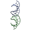
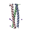
| Title | Radiation damage study on a 16mer DNA segment, structure at 17.7 MGy dose | |||||||||||||||||||||||||||||||
 Components Components | DNA (5'-D(* Keywords KeywordsDNA / radiation damage / global damage / specific damage | Function / homology | DNA / DNA (> 10) |  Function and homology information Function and homology informationBiological species | synthetic construct (others) | Method |  X-RAY DIFFRACTION / SYNCHROTRON / FOURIER SYNTHESIS / Resolution: 1.8 Å X-RAY DIFFRACTION / SYNCHROTRON / FOURIER SYNTHESIS / Resolution: 1.8 Å  Authors AuthorsBugris, V. / Harmat, V. / Ferenc, G. / Brockhauser, S. / Carmichael, I. / Garman, E.F. | Funding support | |  Germany, Germany,  Hungary, 2items Hungary, 2items
 CitationJournal: J.Synchrotron Radiat. / Year: 2019 CitationJournal: J.Synchrotron Radiat. / Year: 2019Title: Radiation-damage investigation of a DNA 16-mer. Authors: Bugris, V. / Harmat, V. / Ferenc, G. / Brockhauser, S. / Carmichael, I. / Garman, E.F. History |
|
- Structure visualization
| Structure viewer | Molecule: MolmilJmol/JSmol |
|---|
- Downloads & links
-Download
| PDBx/mmCIF format | 6qt4.cif.gz | 23.7 KB | Display | PDBx/mmCIF format |
|---|---|---|---|---|
| PDB format | pdb6qt4.ent.gz | 13.9 KB | Display | PDB format |
| PDBx/mmJSON format | 6qt4.json.gz | Tree view | PDBx/mmJSON format | |
| Others |  Other downloads Other downloads |
-Validation report
| Summary document | 6qt4_validation.pdf.gz | 372 KB | Display | wwPDB validaton report |
|---|---|---|---|---|
| Full document | 6qt4_full_validation.pdf.gz | 372.5 KB | Display | |
| Data in XML | 6qt4_validation.xml.gz | 3.2 KB | Display | |
| Data in CIF | 6qt4_validation.cif.gz | 3.9 KB | Display | |
| Arichive directory | https://data.pdbj.org/pub/pdb/validation_reports/qt/6qt4ftp://data.pdbj.org/pub/pdb/validation_reports/qt/6qt4 | HTTPS FTP |
-Related structure data
| Related structure data |  6qt1SC  6qt2C  6qt3C  6qt5C  6qt6C  6qt7 S: Starting model for refinement C: citing same article ( |
|---|---|
| Similar structure data |


-Links
 PDBj
PDBj


- Assembly
| Deposited unit | 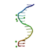
| |||||||||||||||
|---|---|---|---|---|---|---|---|---|---|---|---|---|---|---|---|---|
| 1 | 
| |||||||||||||||
| Unit cell |
| |||||||||||||||
| Components on special symmetry positions |
|
-Components
| #1: DNA chain | Mass: 4898.191 Da / Num. of mol.: 1 / Source method: obtained synthetically Details: Self complementary model DNA sequence derived from kB33 DNA segment. The present sequence is one nucleotide shorter than that of PDB structure 1SGS Source: (synth.) synthetic construct (others) | ||
|---|---|---|---|
| #2: Chemical | ChemComp-CA /   Mass: 40.078 Da / Num. of mol.: 6 / Source method: obtained synthetically / Formula: Ca Mass: 40.078 Da / Num. of mol.: 6 / Source method: obtained synthetically / Formula: Ca#3: Water | ChemComp-HOH / |  Mass: 18.015 Da / Num. of mol.: 38 / Source method: isolated from a natural source / Formula: H2O Mass: 18.015 Da / Num. of mol.: 38 / Source method: isolated from a natural source / Formula: H2O |
-Experimental details
-Experiment
| Experiment | Method: X-RAY DIFFRACTION / Number of used crystals: 1 |
|---|
- Sample preparation
| Crystal | Density Matthews: 2.17 Å3/Da / Density % sol: 43.44 % |
|---|---|
| Crystal grow | Temperature: 292 K / Method: vapor diffusion, hanging drop / pH: 8.6 Details: 2 microliter 1.5 mM DNA solution (5mM HEPES pH 6.6) plus 2 microliter 10 mM HEPES pH 6.6 plus 4 microliter reservoir solution, against a reservoir of 1 ml 34% PEG200, 600 mM CaCl2 and 10 mM HEPES pH 8.6. PH range: 8.6-6.6 |
-Data collection
| Diffraction | Mean temperature: 100 K / Serial crystal experiment: N | |||||||||||||||||||||||||||||||||||||||||||||
|---|---|---|---|---|---|---|---|---|---|---|---|---|---|---|---|---|---|---|---|---|---|---|---|---|---|---|---|---|---|---|---|---|---|---|---|---|---|---|---|---|---|---|---|---|---|---|
| Diffraction source | Source: SYNCHROTRON / Site: Diamond  / Beamline: I24 / Wavelength: 0.9686 Å / Beamline: I24 / Wavelength: 0.9686 Å | |||||||||||||||||||||||||||||||||||||||||||||
| Detector | Type: DECTRIS PILATUS3 6M / Detector: PIXEL / Date: Oct 22, 2018 | |||||||||||||||||||||||||||||||||||||||||||||
| Radiation | Protocol: SINGLE WAVELENGTH / Monochromatic (M) / Laue (L): M / Scattering type: x-ray | |||||||||||||||||||||||||||||||||||||||||||||
| Radiation wavelength | Wavelength: 0.9686 Å / Relative weight: 1 | |||||||||||||||||||||||||||||||||||||||||||||
| Reflection | Resolution: 1.8→31.41 Å / Num. obs: 3898 / % possible obs: 92.2 % / Redundancy: 3 % / CC1/2: 0.995 / Rmerge(I) obs: 0.081 / Rpim(I) all: 0.056 / Rrim(I) all: 0.099 / Net I/σ(I): 3.5 | |||||||||||||||||||||||||||||||||||||||||||||
| Reflection shell | Diffraction-ID: 1
|
- Processing
| Software |
| ||||||||||||||||||||||||||||||||||||||||
|---|---|---|---|---|---|---|---|---|---|---|---|---|---|---|---|---|---|---|---|---|---|---|---|---|---|---|---|---|---|---|---|---|---|---|---|---|---|---|---|---|---|
| Refinement | Method to determine structure: FOURIER SYNTHESIS Starting model: 6QT1 Resolution: 1.8→31.4 Å / Cor.coef. Fo:Fc: 0.965 / Cor.coef. Fo:Fc free: 0.931 / SU B: 6.085 / SU ML: 0.162 / Cross valid method: THROUGHOUT / σ(F): 0 / ESU R: 0.178 / ESU R Free: 0.185 Details: HYDROGENS HAVE BEEN ADDED IN THE RIDING POSITIONS U VALUES : REFINED INDIVIDUALLY. The final model of the lowest dose dataset 6QT1 was further refined against the current dataset collected at higher dose.
| ||||||||||||||||||||||||||||||||||||||||
| Solvent computation | Ion probe radii: 0.8 Å / Shrinkage radii: 0.8 Å / VDW probe radii: 1.2 Å | ||||||||||||||||||||||||||||||||||||||||
| Displacement parameters | Biso max: 126.01 Å2 / Biso mean: 55.095 Å2 / Biso min: 31.39 Å2
| ||||||||||||||||||||||||||||||||||||||||
| Refinement step | Cycle: final / Resolution: 1.8→31.4 Å
| ||||||||||||||||||||||||||||||||||||||||
| Refine LS restraints |
| ||||||||||||||||||||||||||||||||||||||||
| LS refinement shell | Resolution: 1.801→1.847 Å / Rfactor Rfree error: 0 / Total num. of bins used: 20
|
