 Movie
Movie Controller

[English] 日本語
 Yorodumi
- PDB-6qsk: Crystal structure of a nucleotide sugar transporter with bound nu... -
+ Open data

- Basic information
| Entry | Database: PDB / ID: 6qsk | ||||||
|---|---|---|---|---|---|---|---|
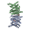
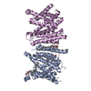
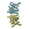
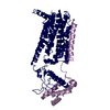
| Title | Crystal structure of a nucleotide sugar transporter with bound nucleotide monophosphate. | ||||||
 Components Components | GDP-mannose transporter 1 | ||||||
 Keywords Keywords | MEMBRANE PROTEIN / Golgi transporter / SLC35 / GDP-mannose transport / glycosylation | ||||||
| Function / homology |  Function and homology information Function and homology informationGDP-mannose transmembrane transporter activity / GDP-mannose transmembrane transport / antiporter activity / cytoplasmic vesicle membrane / Golgi membrane / endoplasmic reticulum membrane / Golgi apparatus / mitochondrion Similarity search - Function | ||||||
| Biological species |  | ||||||
| Method |  X-RAY DIFFRACTION / SYNCHROTRON / MOLECULAR REPLACEMENT / Resolution: 3.394 Å X-RAY DIFFRACTION / SYNCHROTRON / MOLECULAR REPLACEMENT / Resolution: 3.394 Å | ||||||
 Authors Authors | Newstead, S. / Parker, J.L. | ||||||
| Funding support |  United Kingdom, 1items United Kingdom, 1items
| ||||||
 Citation Citation | Journal: Nat Commun / Year: 2019 Title: Structural basis for substrate specificity and regulation of nucleotide sugar transporters in the lipid bilayer. Authors: Parker, J.L. / Corey, R.A. / Stansfeld, P.J. / Newstead, S. | ||||||
| History |
|
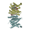
- Structure visualization
| Structure viewer | Molecule: MolmilJmol/JSmol |
|---|
- Downloads & links
-Download
| PDBx/mmCIF format | 6qsk.cif.gz | 463 KB | Display | PDBx/mmCIF format |
|---|---|---|---|---|
| PDB format | pdb6qsk.ent.gz | 379.6 KB | Display | PDB format |
| PDBx/mmJSON format | 6qsk.json.gz | Tree view | PDBx/mmJSON format | |
| Others |  Other downloads Other downloads |
-Validation report
| Arichive directory | https://data.pdbj.org/pub/pdb/validation_reports/qs/6qskftp://data.pdbj.org/pub/pdb/validation_reports/qs/6qsk | HTTPS FTP |
|---|
-Related structure data
| Related structure data |  5ogeS S: Starting model for refinement |
|---|---|
| Similar structure data |


-Links
 PDBj
PDBj


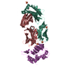
- Assembly
| Deposited unit | 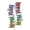
| ||||||||
|---|---|---|---|---|---|---|---|---|---|
| 1 | 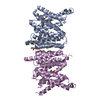
| ||||||||
| 2 | 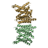
| ||||||||
| 3 | 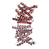
| ||||||||
| 4 | 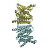
| ||||||||
| Unit cell |
|
-Components
| #1: Protein | Mass: 37044.504 Da / Num. of mol.: 8 Source method: isolated from a genetically manipulated source Source: (gene. exp.) #2: Chemical |   Mass: 356.540 Da / Num. of mol.: 2 / Source method: obtained synthetically / Formula: C21H40O4 Mass: 356.540 Da / Num. of mol.: 2 / Source method: obtained synthetically / Formula: C21H40O4#3: Chemical |   Mass: 363.221 Da / Num. of mol.: 3 / Source method: obtained synthetically / Formula: C10H14N5O8P Mass: 363.221 Da / Num. of mol.: 3 / Source method: obtained synthetically / Formula: C10H14N5O8P#4: Chemical | ChemComp-NA / |   Mass: 22.990 Da / Num. of mol.: 1 / Source method: obtained synthetically / Formula: Na Mass: 22.990 Da / Num. of mol.: 1 / Source method: obtained synthetically / Formula: Na#5: Water | ChemComp-HOH / |  Mass: 18.015 Da / Num. of mol.: 10 / Source method: isolated from a natural source / Formula: H2O Mass: 18.015 Da / Num. of mol.: 10 / Source method: isolated from a natural source / Formula: H2O |
|---|
-Experimental details
-Experiment
| Experiment | Method: X-RAY DIFFRACTION / Number of used crystals: 1 |
|---|
- Sample preparation
| Crystal | Density Matthews: 2.87 Å3/Da / Density % sol: 57.16 % |
|---|---|
| Crystal grow | Temperature: 277 K / Method: lipidic cubic phase / pH: 5 Details: 26 - 30 % (v/v) PEG 400, 0.1 M sodium citrate pH 5.0 and 75 mM sodium chloride or sodium acetate. GMP was soaked in at 20mM overnight. |
-Data collection
| Diffraction | Mean temperature: 83 K / Ambient temp details: 277 / Serial crystal experiment: N |
|---|---|
| Diffraction source | Source: SYNCHROTRON / Site: SOLEIL  / Beamline: PROXIMA 2 / Wavelength: 0.9801 Å / Beamline: PROXIMA 2 / Wavelength: 0.9801 Å |
| Detector | Type: DECTRIS EIGER X 9M / Detector: PIXEL / Date: Sep 30, 2017 |
| Radiation | Protocol: SINGLE WAVELENGTH / Monochromatic (M) / Laue (L): M / Scattering type: x-ray |
| Radiation wavelength | Wavelength: 0.9801 Å / Relative weight: 1 |
| Reflection | Resolution: 3.39→49.43 Å / Num. obs: 44054 / % possible obs: 96.7 % / Redundancy: 2 % / Biso Wilson estimate: 68.12 Å2 / CC1/2: 0.997 / Rmerge(I) obs: 0.1 / Rpim(I) all: 0.09 / Rrim(I) all: 0.135 / Net I/σ(I): 5.4 |
| Reflection shell | Resolution: 3.39→3.52 Å / Redundancy: 2 % / Rmerge(I) obs: 0.68 / Mean I/σ(I) obs: 1.2 / Num. unique obs: 4492 / CC1/2: 0.523 / Rpim(I) all: 0.61 / Rrim(I) all: 0.92 / % possible all: 92.3 |
- Processing
| Software |
| ||||||||||||||||||||||||||||||||||||||||||||||||||||||||||||||||||||||||||||||||||||||||||||||||||||||||||||||||
|---|---|---|---|---|---|---|---|---|---|---|---|---|---|---|---|---|---|---|---|---|---|---|---|---|---|---|---|---|---|---|---|---|---|---|---|---|---|---|---|---|---|---|---|---|---|---|---|---|---|---|---|---|---|---|---|---|---|---|---|---|---|---|---|---|---|---|---|---|---|---|---|---|---|---|---|---|---|---|---|---|---|---|---|---|---|---|---|---|---|---|---|---|---|---|---|---|---|---|---|---|---|---|---|---|---|---|---|---|---|---|---|---|---|
| Refinement | Method to determine structure: MOLECULAR REPLACEMENT Starting model: 5OGE Resolution: 3.394→49.428 Å / SU ML: 0.63 / Cross valid method: THROUGHOUT / σ(F): 1.96 / Phase error: 36.65 / Stereochemistry target values: ML
| ||||||||||||||||||||||||||||||||||||||||||||||||||||||||||||||||||||||||||||||||||||||||||||||||||||||||||||||||
| Solvent computation | Shrinkage radii: 0.9 Å / VDW probe radii: 1.11 Å / Solvent model: FLAT BULK SOLVENT MODEL | ||||||||||||||||||||||||||||||||||||||||||||||||||||||||||||||||||||||||||||||||||||||||||||||||||||||||||||||||
| Refinement step | Cycle: LAST / Resolution: 3.394→49.428 Å
| ||||||||||||||||||||||||||||||||||||||||||||||||||||||||||||||||||||||||||||||||||||||||||||||||||||||||||||||||
| Refine LS restraints |
| ||||||||||||||||||||||||||||||||||||||||||||||||||||||||||||||||||||||||||||||||||||||||||||||||||||||||||||||||
| LS refinement shell |
|
