 Movie
Movie Controller

[English] 日本語
 Yorodumi
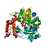
- PDB-6qbw: Structure of the HTLV-2 integrase catalytic core domain in comple... -
+ Open data

- Basic information
| Entry | Database: PDB / ID: 6qbw | ||||||
|---|---|---|---|---|---|---|---|
| Title | Structure of the HTLV-2 integrase catalytic core domain in complex with calcium | ||||||
 Components Components | integrase | ||||||
 Keywords Keywords | TRANSFERASE / retrovirus / deltaretrovirus / integration / strand-transfer / nucleotidyltransferase | ||||||
| Function / homology |  Function and homology information Function and homology informationHydrolases; Acting on peptide bonds (peptidases); Aspartic endopeptidases / ribonuclease H / DNA integration / viral genome integration into host DNA / establishment of integrated proviral latency / RNA-directed DNA polymerase / RNA stem-loop binding / RNA-directed DNA polymerase activity / RNA-DNA hybrid ribonuclease activity / Transferases; Transferring phosphorus-containing groups; Nucleotidyltransferases ...Hydrolases; Acting on peptide bonds (peptidases); Aspartic endopeptidases / ribonuclease H / DNA integration / viral genome integration into host DNA / establishment of integrated proviral latency / RNA-directed DNA polymerase / RNA stem-loop binding / RNA-directed DNA polymerase activity / RNA-DNA hybrid ribonuclease activity / Transferases; Transferring phosphorus-containing groups; Nucleotidyltransferases / viral nucleocapsid / DNA recombination / DNA-directed DNA polymerase / aspartic-type endopeptidase activity / Hydrolases; Acting on ester bonds / DNA-directed DNA polymerase activity / symbiont-mediated suppression of host gene expression / viral translational frameshifting / symbiont entry into host cell / structural molecule activity / proteolysis / DNA binding / zinc ion binding Similarity search - Function | ||||||
| Biological species |  Human T-cell leukemia virus 2 Human T-cell leukemia virus 2 | ||||||
| Method |  X-RAY DIFFRACTION / SYNCHROTRON / MOLECULAR REPLACEMENT / Resolution: 2.4 Å X-RAY DIFFRACTION / SYNCHROTRON / MOLECULAR REPLACEMENT / Resolution: 2.4 Å | ||||||
 Authors Authors | Barski, M.S. / Maertens, G.N. | ||||||
| Funding support |  United Kingdom, 1items United Kingdom, 1items
| ||||||
 Citation Citation | Journal: Nat Commun / Year: 2020 Title: Cryo-EM structure of the deltaretroviral intasome in complex with the PP2A regulatory subunit B56γ. Authors: Michał S Barski / Jordan J Minnell / Zuzana Hodakova / Valerie E Pye / Andrea Nans / Peter Cherepanov / Goedele N Maertens / Abstract: Human T-cell lymphotropic virus type 1 (HTLV-1) is a deltaretrovirus and the most oncogenic pathogen. Many of the ~20 million HTLV-1 infected people will develop severe leukaemia or an ALS-like motor ...Human T-cell lymphotropic virus type 1 (HTLV-1) is a deltaretrovirus and the most oncogenic pathogen. Many of the ~20 million HTLV-1 infected people will develop severe leukaemia or an ALS-like motor disease, unless a therapy becomes available. A key step in the establishment of infection is the integration of viral genetic material into the host genome, catalysed by the retroviral integrase (IN) enzyme. Here, we use X-ray crystallography and single-particle cryo-electron microscopy to determine the structure of the functional deltaretroviral IN assembled on viral DNA ends and bound to the B56γ subunit of its human host factor, protein phosphatase 2 A. The structure reveals a tetrameric IN assembly bound to two molecules of the phosphatase via a conserved short linear motif. Insight into the deltaretroviral intasome and its interaction with the host will be crucial for understanding the pattern of integration events in infected individuals and therefore bears important clinical implications. | ||||||
| History |
|
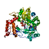
- Structure visualization
| Structure viewer | Molecule: MolmilJmol/JSmol |
|---|
- Downloads & links
-Download
| PDBx/mmCIF format | 6qbw.cif.gz | 44.9 KB | Display | PDBx/mmCIF format |
|---|---|---|---|---|
| PDB format | pdb6qbw.ent.gz | 29.7 KB | Display | PDB format |
| PDBx/mmJSON format | 6qbw.json.gz | Tree view | PDBx/mmJSON format | |
| Others |  Other downloads Other downloads |
-Validation report
| Arichive directory | https://data.pdbj.org/pub/pdb/validation_reports/qb/6qbwftp://data.pdbj.org/pub/pdb/validation_reports/qb/6qbw | HTTPS FTP |
|---|
-Related structure data
| Related structure data |  6qbtSC 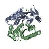 6qbvC  6tjuC  6toqC 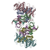 7pelC S: Starting model for refinement C: citing same article ( |
|---|---|
| Similar structure data |

-Links
 PDBj
PDBj

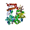
- Assembly
| Deposited unit | 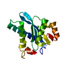
| ||||||||
|---|---|---|---|---|---|---|---|---|---|
| 1 | 
| ||||||||
| Unit cell |
|
-Components
| #1: Protein | Mass: 19178.967 Da / Num. of mol.: 1 Source method: isolated from a genetically manipulated source Source: (gene. exp.) Human T-cell leukemia virus 2 / Gene: pol / Production host:  |
|---|---|
| #2: Chemical | ChemComp-CA /   Mass: 40.078 Da / Num. of mol.: 1 / Source method: obtained synthetically / Formula: Ca Mass: 40.078 Da / Num. of mol.: 1 / Source method: obtained synthetically / Formula: Ca |
| #3: Water | ChemComp-HOH /  Mass: 18.015 Da / Num. of mol.: 28 / Source method: isolated from a natural source / Formula: H2O Mass: 18.015 Da / Num. of mol.: 28 / Source method: isolated from a natural source / Formula: H2O |
| Has protein modification | Y |
-Experimental details
-Experiment
| Experiment | Method: X-RAY DIFFRACTION / Number of used crystals: 1 |
|---|
- Sample preparation
| Crystal | Density Matthews: 3.06 Å3/Da / Density % sol: 65.93 % |
|---|---|
| Crystal grow | Temperature: 291 K / Method: vapor diffusion, hanging drop / pH: 8 Details: 100 mM Tris pH 8, 18% PEG 6000, 250 calcium chloride dihydrate |
-Data collection
| Diffraction | Mean temperature: 291 K / Serial crystal experiment: N |
|---|---|
| Diffraction source | Source: SYNCHROTRON / Site: Diamond / Beamline: I04 / Wavelength: 0.9795 Å |
| Detector | Type: DECTRIS PILATUS 6M-F / Detector: PIXEL / Date: May 11, 2018 |
| Radiation | Protocol: SINGLE WAVELENGTH / Monochromatic (M) / Laue (L): M / Scattering type: x-ray |
| Radiation wavelength | Wavelength: 0.9795 Å / Relative weight: 1 |
| Reflection | Resolution: 2.4→50.12 Å / Num. obs: 9932 / % possible obs: 100 % / Redundancy: 1.8 % / Rmerge(I) obs: 0.028 / Net I/σ(I): 13.8 |
| Reflection shell | Resolution: 2.4→2.49 Å / Redundancy: 1.9 % / Rmerge(I) obs: 0.31 / Mean I/σ(I) obs: 1.8 / Num. unique obs: 1872 / % possible all: 100 |
- Processing
| Software |
| ||||||||||||||||
|---|---|---|---|---|---|---|---|---|---|---|---|---|---|---|---|---|---|
| Refinement | Method to determine structure: MOLECULAR REPLACEMENT Starting model: 6QBT Resolution: 2.4→50.12 Å / Cross valid method: FREE R-VALUE
| ||||||||||||||||
| Solvent computation | Ion probe radii: 0.9 Å / VDW probe radii: 1.2 Å | ||||||||||||||||
| Displacement parameters | Biso mean: 53.312 Å2 | ||||||||||||||||
| Refinement step | Cycle: LAST / Resolution: 2.4→50.12 Å
|
