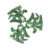
negative regulation of chemokine (C-C motif) ligand 4 production / negative regulation of activated CD8-positive, alpha-beta T cell apoptotic process / negative regulation of defense response to bacterium / negative regulation of type 2 immune response / negative regulation of macrophage inflammatory protein 1 alpha production / negative regulation of interleukin-13 production / negative regulation of chemokine (C-C motif) ligand 5 production / regulation of interleukin-1 beta production / Urea cycle / arginine metabolic process ...negative regulation of chemokine (C-C motif) ligand 4 production / negative regulation of activated CD8-positive, alpha-beta T cell apoptotic process / negative regulation of defense response to bacterium / negative regulation of type 2 immune response / negative regulation of macrophage inflammatory protein 1 alpha production / negative regulation of interleukin-13 production / negative regulation of chemokine (C-C motif) ligand 5 production / regulation of interleukin-1 beta production / Urea cycle / arginine metabolic process / arginase / arginase activity / urea cycle / negative regulation of CD4-positive, alpha-beta T cell proliferation / negative regulation of interleukin-17 production / ureteric bud development / regulation of reactive oxygen species biosynthetic process / negative regulation of tumor necrosis factor production / striated muscle contraction / nitric oxide biosynthetic process / Mitochondrial protein degradation / positive regulation of cellular senescence / manganese ion binding / adaptive immune response / mitochondrial matrix / innate immune response / mitochondrion / cytoplasm Similarity search - Function
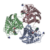
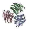
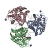
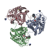
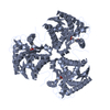
Arginase / Ureohydrolase domain / Ureohydrolase, manganese-binding site / Arginase family signature. / Ureohydrolase / Arginase family / Arginase family profile. / Arginase; Chain A / Ureohydrolase domain superfamily / 3-Layer(aba) Sandwich / Alpha Beta Similarity search - Domain/homology
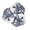
Mass: 18.015 Da / Num. of mol.: 824 / Source method: isolated from a natural source / Formula: H2O
-
Experimental details
-
Experiment
Experiment
Method: X-RAY DIFFRACTION / Number of used crystals: 1
-
Sample preparation
Crystal
Density Matthews: 3.27 Å3/Da / Density % sol: 62.42 %
Crystal grow
Temperature: 293 K / Method: vapor diffusion, hanging drop / pH: 7 Details: CRYSTALS IN CONDITION H3 FROM SILVER BULLET SCREEN (HAMPTON RESEARCH) USING TACSIMATE
-
Data collection
Diffraction
Mean temperature: 100 K / Serial crystal experiment: N
In the structure databanks used in Yorodumi, some data are registered as the other names, "COVID-19 virus" and "2019-nCoV". Here are the details of the virus and the list of structure data.
Jan 31, 2019. EMDB accession codes are about to change! (news from PDBe EMDB page)
EMDB accession codes are about to change! (news from PDBe EMDB page)
The allocation of 4 digits for EMDB accession codes will soon come to an end. Whilst these codes will remain in use, new EMDB accession codes will include an additional digit and will expand incrementally as the available range of codes is exhausted. The current 4-digit format prefixed with “EMD-” (i.e. EMD-XXXX) will advance to a 5-digit format (i.e. EMD-XXXXX), and so on. It is currently estimated that the 4-digit codes will be depleted around Spring 2019, at which point the 5-digit format will come into force.
The EM Navigator/Yorodumi systems omit the EMD- prefix.
Related info.:Q: What is EMD? / ID/Accession-code notation in Yorodumi/EM Navigator
Yorodumi is a browser for structure data from EMDB, PDB, SASBDB, etc.
This page is also the successor to EM Navigator detail page, and also detail information page/front-end page for Omokage search.
The word "yorodu" (or yorozu) is an old Japanese word meaning "ten thousand". "mi" (miru) is to see.
Related info.:EMDB / PDB / SASBDB / Comparison of 3 databanks / Yorodumi Search / Aug 31, 2016. New EM Navigator & Yorodumi / Yorodumi Papers / Jmol/JSmol / Function and homology information / Changes in new EM Navigator and Yorodumi
 Movie
Movie Controller


 Open data
 Basic information
 Components
 Keywords
 Function and homology information
 Homo sapiens (human)
 X-RAY DIFFRACTION /
  Authors
 Citation
 Structure visualization
 Downloads & links
 Other downloads


 PDBj
PDBj


 Assembly
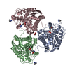


 Mass: 54.938 Da / Num. of mol.: 6 / Source method: obtained synthetically / Formula: Mn
 Mass: 120.152 Da / Num. of mol.: 3 / Source method: obtained synthetically / Formula: C7H8N2
 Mass: 78.133 Da / Num. of mol.: 3 / Source method: obtained synthetically / Formula: C2H6OS
 Mass: 233.050 Da / Num. of mol.: 3 / Source method: obtained synthetically / Formula: C8H18BN2O5 / Feature type: SUBJECT OF INVESTIGATION
 Mass: 62.068 Da / Num. of mol.: 1 / Source method: obtained synthetically / Formula: C2H6O2
 Sample preparation
 Processing