 ムービー
ムービー コントローラー


+ データを開く

- 基本情報
基本情報
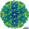
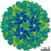
| 登録情報 | データベース: PDB / ID: 6q1f | |||||||||||||||
|---|---|---|---|---|---|---|---|---|---|---|---|---|---|---|---|---|
| タイトル | Atomic structure of the Human Herpesvirus 6B Capsid and Capsid-Associated Tegument Complexes | |||||||||||||||
 要素 要素 |
| |||||||||||||||
 キーワード キーワード | VIRUS / beta-herpesvirus / HHV-6B / murine cytomegalovirus / human cytomegalovirus / pp150 / pU11 / pUL32 / pM32 / pU14 | |||||||||||||||
| 機能・相同性 |  機能・相同性情報 機能・相同性情報T=16 icosahedral viral capsid / viral tegument / viral capsid assembly / viral process / viral capsid / host cell nucleus / structural molecule activity / DNA binding 類似検索 - 分子機能 | |||||||||||||||
| 生物種 |  Human herpesvirus 6B (ヘルペスウイルス) Human herpesvirus 6B (ヘルペスウイルス) | |||||||||||||||
| 手法 | 電子顕微鏡法 / 単粒子再構成法 / クライオ電子顕微鏡法 / 解像度: 9 Å | |||||||||||||||
 データ登録者 データ登録者 | Zhang, Y.B. / Liu, W. / Li, Z.H. / Kumar, V. / Alvarez-Cabrera, A.L. / Leibovitch, E. / Cui, Y.X. / Mei, Y. / Bi, G.Q. / Jacobson, S. / Zhou, Z.H. | |||||||||||||||
| 資金援助 |  米国, 4件 米国, 4件
| |||||||||||||||
 引用 引用 | ジャーナル: Nat Commun / 年: 2019 タイトル: Atomic structure of the human herpesvirus 6B capsid and capsid-associated tegument complexes. 著者: Yibo Zhang / Wei Liu / Zihang Li / Vinay Kumar / Ana L Alvarez-Cabrera / Emily C Leibovitch / Yanxiang Cui / Ye Mei / Guo-Qiang Bi / Steve Jacobson / Z Hong Zhou /  要旨: Human herpesvirus 6B (HHV-6B) belongs to the β-herpesvirus subfamily of the Herpesviridae. To understand capsid assembly and capsid-tegument interactions, here we report atomic structures of HHV-6B ...Human herpesvirus 6B (HHV-6B) belongs to the β-herpesvirus subfamily of the Herpesviridae. To understand capsid assembly and capsid-tegument interactions, here we report atomic structures of HHV-6B capsid and capsid-associated tegument complex (CATC) obtained by cryoEM and sub-particle reconstruction. Compared to other β-herpesviruses, HHV-6B exhibits high similarity in capsid structure but organizational differences in its CATC (pU11 tetramer). 180 "VΛ"-shaped CATCs are observed in HHV-6B, distinguishing from the 255 "Λ"-shaped dimeric CATCs observed in murine cytomegalovirus and the 310 "Δ"-shaped CATCs in human cytomegalovirus. This trend in CATC quantity correlates with the increasing genomes sizes of these β-herpesviruses. Incompatible distances revealed by the atomic structures rationalize the lack of CATC's binding to triplexes Ta, Tc, and Tf in HHV-6B. Our results offer insights into HHV-6B capsid assembly and the roles of its tegument proteins, including not only the β-herpesvirus-specific pU11 and pU14, but also those conserved across all subfamilies of Herpesviridae. | |||||||||||||||
| 履歴 |
|
- 構造の表示
構造の表示
| ムービー |
ムービービューア |
|---|---|
| 構造ビューア | 分子: MolmilJmol/JSmol |
- ダウンロードとリンク
-ダウンロード
| PDBx/mmCIF形式 | 6q1f.cif.gz | 5.1 MB | 表示 | PDBx/mmCIF形式 |
|---|---|---|---|---|
| PDB形式 | pdb6q1f.ent.gz | 表示 | PDB形式 | |
| PDBx/mmJSON形式 | 6q1f.json.gz | ツリー表示 | PDBx/mmJSON形式 | |
| その他 |  その他のダウンロード その他のダウンロード |
-検証レポート
| アーカイブディレクトリ | https://data.pdbj.org/pub/pdb/validation_reports/q1/6q1fftp://data.pdbj.org/pub/pdb/validation_reports/q1/6q1f | HTTPS FTP |
|---|
-関連構造データ


-リンク
 PDBj
PDBj
- 集合体
集合体
| 登録構造単位 | 
|
|---|---|
| 1 | x 60
|
| 2 |
|
| 3 | x 5
|
| 4 | x 6
|
| 5 | 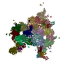
|
| 対称性 | 点対称性: (シェーンフリース記号: I (正20面体型対称)) |
-要素
| #1: タンパク質 | 分子量: 152260.047 Da / 分子数: 16 / 由来タイプ: 天然 由来: (天然) Human herpesvirus 6B (strain Z29) (ヘルペスウイルス)株: Z29 / 参照: UniProt: Q9QJ26 #2: タンパク質 | 分子量: 95738.125 Da / 分子数: 12 / 由来タイプ: 天然 由来: (天然) Human herpesvirus 6B (strain Z29) (ヘルペスウイルス)株: Z29 / 参照: UniProt: Q69535 #3: タンパク質 | 分子量: 9827.329 Da / 分子数: 16 / 由来タイプ: 天然 由来: (天然) Human herpesvirus 6B (strain Z29) (ヘルペスウイルス)株: Z29 / 参照: UniProt: Q9WT32 #4: タンパク質 | 分子量: 34162.508 Da / 分子数: 5 / 由来タイプ: 天然 由来: (天然) Human herpesvirus 6B (strain Z29) (ヘルペスウイルス)株: Z29 / 参照: UniProt: Q9WT35 #5: タンパク質 | 分子量: 33514.332 Da / 分子数: 10 / 由来タイプ: 天然 由来: (天然) Human herpesvirus 6B (strain Z29) (ヘルペスウイルス)株: Z29 / 参照: UniProt: Q9QJ27 |
|---|
-実験情報
-実験
| 実験 | 手法: 電子顕微鏡法 |
|---|---|
| EM実験 | 試料の集合状態: PARTICLE / 3次元再構成法: 単粒子再構成法 |
- 試料調製
試料調製
| 構成要素 | 名称: Human herpesvirus 6 strain Z29 / タイプ: VIRUS / Entity ID: all / 由来: NATURAL |
|---|---|
| 分子量 | 実験値: NO |
| 由来(天然) | 生物種: Human herpesvirus 6 strain Z29 (ヘルペスウイルス) 株: Z29 |
| ウイルスについての詳細 | 中空か: NO / エンベロープを持つか: YES / 単離: STRAIN / タイプ: VIRION |
| 緩衝液 | pH: 7.4 / 詳細: PBS buffer, pH 7.4 |
| 試料 | 包埋: NO / シャドウイング: NO / 染色: NO / 凍結: YES |
| 試料支持 | グリッドの材料: COPPER / グリッドのサイズ: 200 divisions/in. |
| 急速凍結 | 装置: HOMEMADE PLUNGER / 凍結剤: ETHANE / 詳細: The grids were manually plunged into the ethane. |
- 電子顕微鏡撮影
| 実験機器 |  モデル: Titan Krios / 画像提供: FEI Company |
|---|---|
| 顕微鏡 | モデル: FEI TITAN KRIOS |
| 電子銃 | 電子線源:  FIELD EMISSION GUN / 加速電圧: 300 kV / 照射モード: FLOOD BEAM FIELD EMISSION GUN / 加速電圧: 300 kV / 照射モード: FLOOD BEAM |
| 電子レンズ | モード: BRIGHT FIELD / 倍率(公称値): 64000 X / 倍率(補正後): 64000 X / 最大 デフォーカス(公称値): 3200 nm / 最小 デフォーカス(公称値): 2200 nm / Calibrated defocus min: 2200 nm / 最大 デフォーカス(補正後): 3200 nm / Cs: 2.7 mm / アライメント法: COMA FREE |
| 試料ホルダ | 凍結剤: NITROGEN 試料ホルダーモデル: FEI TITAN KRIOS AUTOGRID HOLDER |
| 撮影 | 電子線照射量: 23 e/Å2 フィルム・検出器のモデル: GATAN K2 SUMMIT (4k x 4k) 実像数: 4828 |
- 解析
解析
| EMソフトウェア |
| ||||||||||||
|---|---|---|---|---|---|---|---|---|---|---|---|---|---|
| CTF補正 | タイプ: PHASE FLIPPING AND AMPLITUDE CORRECTION | ||||||||||||
| 粒子像の選択 | 選択した粒子像数: 7430 | ||||||||||||
| 対称性 | 点対称性: I (正20面体型対称) | ||||||||||||
| 3次元再構成 | 解像度: 9 Å / 解像度の算出法: FSC 0.143 CUT-OFF / 粒子像の数: 6443 / 詳細: bin4 reconstrction / 対称性のタイプ: POINT | ||||||||||||
| 原子モデル構築 | プロトコル: OTHER |
