 Movie
Movie Controller

[English] 日本語
 Yorodumi
- PDB-6q0g: Crystal structure of ligand-binding domain of Pseudomonas fluores... -
+ Open data

- Basic information
| Entry | Database: PDB / ID: 6q0g | ||||||
|---|---|---|---|---|---|---|---|
| Title | Crystal structure of ligand-binding domain of Pseudomonas fluorescens chemoreceptor CtaA in complex with L-proline | ||||||
 Components Components | Putative methyl-accepting chemotaxis protein | ||||||
 Keywords Keywords | SIGNALING PROTEIN / Bacterial chemotaxis / chemoreceptor / double Cache / ligand binding domain | ||||||
| Function / homology |  Function and homology information Function and homology informationamino acid binding / response to amino acid / chemotaxis / transmembrane signaling receptor activity / signal transduction / plasma membrane Similarity search - Function | ||||||
| Biological species |  Pseudomonas fluorescens (bacteria) Pseudomonas fluorescens (bacteria) | ||||||
| Method |  X-RAY DIFFRACTION / SYNCHROTRON / MOLECULAR REPLACEMENT / Resolution: 2 Å X-RAY DIFFRACTION / SYNCHROTRON / MOLECULAR REPLACEMENT / Resolution: 2 Å | ||||||
 Authors Authors | Ud-Din, I.A. / Khan, M.F. / Roujeinikova, A. | ||||||
| Funding support |  Australia, 1items Australia, 1items
| ||||||
 Citation Citation | Journal: Mol.Plant Microbe Interact. / Year: 2020 Title: Broad Specificity of Amino Acid Chemoreceptor CtaA ofPseudomonas fluorescensIs Afforded by Plasticity of Its Amphipathic Ligand-Binding Pocket. Authors: Ud-Din, A.I.M.S. / Khan, M.F. / Roujeinikova, A. | ||||||
| History |
|
- Structure visualization
| Structure viewer | Molecule: MolmilJmol/JSmol |
|---|
- Downloads & links
-Download
| PDBx/mmCIF format | 6q0g.cif.gz | 62.3 KB | Display | PDBx/mmCIF format |
|---|---|---|---|---|
| PDB format | pdb6q0g.ent.gz | 42.4 KB | Display | PDB format |
| PDBx/mmJSON format | 6q0g.json.gz | Tree view | PDBx/mmJSON format | |
| Others |  Other downloads Other downloads |
-Validation report
| Arichive directory | https://data.pdbj.org/pub/pdb/validation_reports/q0/6q0gftp://data.pdbj.org/pub/pdb/validation_reports/q0/6q0g | HTTPS FTP |
|---|
-Related structure data
| Related structure data |  6pxyC  6py3C 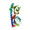 6py4C 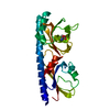 6py5C 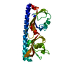 6pyiC  6q0fC 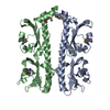 3c8cS S: Starting model for refinement C: citing same article ( |
|---|---|
| Similar structure data |


-Links
 PDBj
PDBj


- Assembly
| Deposited unit | 
| |||||||||
|---|---|---|---|---|---|---|---|---|---|---|
| 1 |
| |||||||||
| Unit cell |
| |||||||||
| Components on special symmetry positions |
|
-Components
| #1: Protein | Mass: 27055.496 Da / Num. of mol.: 1 Source method: isolated from a genetically manipulated source Source: (gene. exp.) Pseudomonas fluorescens (strain Pf0-1) (bacteria)Strain: Pf0-1 / Gene: Pfl01_4431 / Production host: | ||||
|---|---|---|---|---|---|
| #2: Chemical | ChemComp-PRO /   Type: L-peptide linking / Mass: 115.130 Da / Num. of mol.: 1 / Source method: obtained synthetically / Formula: C5H9NO2 / Feature type: SUBJECT OF INVESTIGATION Type: L-peptide linking / Mass: 115.130 Da / Num. of mol.: 1 / Source method: obtained synthetically / Formula: C5H9NO2 / Feature type: SUBJECT OF INVESTIGATION | ||||
| #3: Chemical |   Mass: 35.453 Da / Num. of mol.: 2 / Source method: isolated from a natural source / Formula: Cl Mass: 35.453 Da / Num. of mol.: 2 / Source method: isolated from a natural source / Formula: Cl#4: Water | ChemComp-HOH / |  Mass: 18.015 Da / Num. of mol.: 193 / Source method: isolated from a natural source / Formula: H2O Mass: 18.015 Da / Num. of mol.: 193 / Source method: isolated from a natural source / Formula: H2OHas ligand of interest | Y | |
-Experimental details
-Experiment
| Experiment | Method: X-RAY DIFFRACTION / Number of used crystals: 1 |
|---|
- Sample preparation
| Crystal | Density Matthews: 2.3 Å3/Da / Density % sol: 46.5 % |
|---|---|
| Crystal grow | Temperature: 298 K / Method: vapor diffusion, hanging drop / Details: Ammonium sulfate and Tris-HCl |
-Data collection
| Diffraction | Mean temperature: 100 K / Serial crystal experiment: N | |||||||||||||||||||||||||||||||||||||||||||||||||||||||||||||||||||||||||||||||||||||||||||||||||||||||||||||||||||||||||
|---|---|---|---|---|---|---|---|---|---|---|---|---|---|---|---|---|---|---|---|---|---|---|---|---|---|---|---|---|---|---|---|---|---|---|---|---|---|---|---|---|---|---|---|---|---|---|---|---|---|---|---|---|---|---|---|---|---|---|---|---|---|---|---|---|---|---|---|---|---|---|---|---|---|---|---|---|---|---|---|---|---|---|---|---|---|---|---|---|---|---|---|---|---|---|---|---|---|---|---|---|---|---|---|---|---|---|---|---|---|---|---|---|---|---|---|---|---|---|---|---|---|---|
| Diffraction source | Source: SYNCHROTRON / Site: Australian Synchrotron / Beamline: MX1 / Wavelength: 1 Å | |||||||||||||||||||||||||||||||||||||||||||||||||||||||||||||||||||||||||||||||||||||||||||||||||||||||||||||||||||||||||
| Detector | Type: ADSC QUANTUM 210r / Detector: CCD / Date: Oct 10, 2017 | |||||||||||||||||||||||||||||||||||||||||||||||||||||||||||||||||||||||||||||||||||||||||||||||||||||||||||||||||||||||||
| Radiation | Protocol: SINGLE WAVELENGTH / Monochromatic (M) / Laue (L): M / Scattering type: x-ray | |||||||||||||||||||||||||||||||||||||||||||||||||||||||||||||||||||||||||||||||||||||||||||||||||||||||||||||||||||||||||
| Radiation wavelength | Wavelength: 1 Å / Relative weight: 1 | |||||||||||||||||||||||||||||||||||||||||||||||||||||||||||||||||||||||||||||||||||||||||||||||||||||||||||||||||||||||||
| Reflection | Resolution: 2→60.478 Å / Num. obs: 17230 / % possible obs: 100 % / Redundancy: 6.9 % / Biso Wilson estimate: 27.55 Å2 / Rpim(I) all: 0.038 / Rrim(I) all: 0.1 / Rsym value: 0.092 / Net I/av σ(I): 4.4 / Net I/σ(I): 11.7 / Num. measured all: 118988 | |||||||||||||||||||||||||||||||||||||||||||||||||||||||||||||||||||||||||||||||||||||||||||||||||||||||||||||||||||||||||
| Reflection shell | Diffraction-ID: 1
|
- Processing
| Software |
| ||||||||||||||||||||||||||||||
|---|---|---|---|---|---|---|---|---|---|---|---|---|---|---|---|---|---|---|---|---|---|---|---|---|---|---|---|---|---|---|---|
| Refinement | Method to determine structure: MOLECULAR REPLACEMENT Starting model: 3C8C Resolution: 2→35.953 Å / SU ML: 0.18 / Cross valid method: THROUGHOUT / σ(F): 1.34 / Phase error: 29.21 / Stereochemistry target values: ML
| ||||||||||||||||||||||||||||||
| Solvent computation | Shrinkage radii: 0.9 Å / VDW probe radii: 1.11 Å / Solvent model: FLAT BULK SOLVENT MODEL | ||||||||||||||||||||||||||||||
| Displacement parameters | Biso max: 95.45 Å2 / Biso mean: 39.4568 Å2 / Biso min: 14.87 Å2 | ||||||||||||||||||||||||||||||
| Refinement step | Cycle: final / Resolution: 2→35.953 Å
| ||||||||||||||||||||||||||||||
| Refine LS restraints |
| ||||||||||||||||||||||||||||||
| LS refinement shell | Refine-ID: X-RAY DIFFRACTION / Rfactor Rfree error: 0 / % reflection obs: 100 %
|
