Mass: 18.015 Da / Num. of mol.: 321 / Source method: isolated from a natural source / Formula: H2O
-
Details
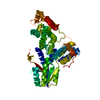
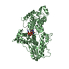
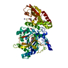
Has ligand of interest
Y
-
Experimental details
-
Experiment
Experiment
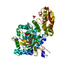
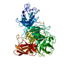
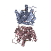
Method: X-RAY DIFFRACTION / Number of used crystals: 1
-
Sample preparation
Crystal
Density Matthews: 2.19 Å3/Da / Density % sol: 43.74 %
Crystal grow
Temperature: 294.15 K / Method: vapor diffusion, hanging drop Details: Crystals were obtained at 21C in dark from a 1:1 mixture of protein (10-12 mg/mL) and a reservoir solution consisting of 5% PEG 6000 and 0.1 M HEPES pH 7.1. Crystal reached maximum size ...Details: Crystals were obtained at 21C in dark from a 1:1 mixture of protein (10-12 mg/mL) and a reservoir solution consisting of 5% PEG 6000 and 0.1 M HEPES pH 7.1. Crystal reached maximum size after 4-5 days and were harvested at that time in the dark. Crystals were transferred to cryoprotectant solution consisting of 5% PEG 6000, 0.1 M HEPES pH 7, 2 mM DTT and either 30% P400 or 30% glycerol. Crystals were flash-frozen in liquid nitrogen in the dark
-
Data collection
Diffraction
Mean temperature: 100 K / Serial crystal experiment: N
In the structure databanks used in Yorodumi, some data are registered as the other names, "COVID-19 virus" and "2019-nCoV". Here are the details of the virus and the list of structure data.
Jan 31, 2019. EMDB accession codes are about to change! (news from PDBe EMDB page)
EMDB accession codes are about to change! (news from PDBe EMDB page)
The allocation of 4 digits for EMDB accession codes will soon come to an end. Whilst these codes will remain in use, new EMDB accession codes will include an additional digit and will expand incrementally as the available range of codes is exhausted. The current 4-digit format prefixed with “EMD-” (i.e. EMD-XXXX) will advance to a 5-digit format (i.e. EMD-XXXXX), and so on. It is currently estimated that the 4-digit codes will be depleted around Spring 2019, at which point the 5-digit format will come into force.
The EM Navigator/Yorodumi systems omit the EMD- prefix.
Related info.:Q: What is EMD? / ID/Accession-code notation in Yorodumi/EM Navigator
Yorodumi is a browser for structure data from EMDB, PDB, SASBDB, etc.
This page is also the successor to EM Navigator detail page, and also detail information page/front-end page for Omokage search.
The word "yorodu" (or yorozu) is an old Japanese word meaning "ten thousand". "mi" (miru) is to see.
Related info.:EMDB / PDB / SASBDB / Comparison of 3 databanks / Yorodumi Search / Aug 31, 2016. New EM Navigator & Yorodumi / Yorodumi Papers / Jmol/JSmol / Function and homology information / Changes in new EM Navigator and Yorodumi
 Movie
Movie Controller


 Open data
 Basic information
 Components
 Keywords
 Function and homology information
 X-RAY DIFFRACTION /
  Authors
 United States, European Union,
  Germany, 10items
  Citation
 Structure visualization
 Downloads & links
 Other downloads
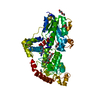


 PDBj
PDBj

 Assembly
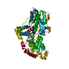

 Spodoptera frugiperda (fall armyworm) / References: UniProt: A0A2I0LZR8


 Mass: 785.550 Da / Num. of mol.: 1 / Source method: obtained synthetically / Formula: C27H33N9O15P2 / Feature type: SUBJECT OF INVESTIGATION / Comment: FAD*YM
 Mass: 150.173 Da / Num. of mol.: 1 / Source method: obtained synthetically / Formula: C6H14O4
 Mass: 62.068 Da / Num. of mol.: 1 / Source method: obtained synthetically / Formula: C2H6O2
 Mass: 106.120 Da / Num. of mol.: 4 / Source method: obtained synthetically / Formula: C4H10O3
 Mass: 92.094 Da / Num. of mol.: 1 / Source method: obtained synthetically / Formula: C3H8O3
 Sample preparation
 Processing