 Movie
Movie Controller

[English] 日本語
 Yorodumi
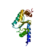
- PDB-6pq7: Structure of the iMango-III fluorescent aptamer at room temperature. -
+ Open data

- Basic information
| Entry | Database: PDB / ID: 6pq7 | ||||||
|---|---|---|---|---|---|---|---|
| Title | Structure of the iMango-III fluorescent aptamer at room temperature. | ||||||
 Components Components | RNA (37-MER) | ||||||
 Keywords Keywords | RNA / fluorescent / aptamer / XFELS | ||||||
| Function / homology | : / Chem-OXV / RNA / RNA (> 10) Function and homology information Function and homology information | ||||||
| Biological species | synthetic construct (others) | ||||||
| Method |  X-RAY DIFFRACTION / FREE ELECTRON LASER / MOLECULAR REPLACEMENT / Resolution: 3 Å X-RAY DIFFRACTION / FREE ELECTRON LASER / MOLECULAR REPLACEMENT / Resolution: 3 Å | ||||||
 Authors Authors | Trachman III, R.J. / Ferre-D'Amare, A.R. | ||||||
 Citation Citation | Journal: Acta Crystallogr.,Sect.F / Year: 2019 Title: Co-crystal structure of the iMango-III fluorescent RNA aptamer using an X-ray free-electron laser. Authors: Trachman III, R.J. / Stagno, J.R. / Conrad, C. / Jones, C.P. / Fischer, P. / Meents, A. / Wang, Y.X. / Ferre-D'Amare, A.R. #1: Journal: Acta Crystallogr D Biol Crystallogr / Year: 2010 Title: PHENIX: a comprehensive Python-based system for macromolecular structure solution. Authors: Paul D Adams / Pavel V Afonine / Gábor Bunkóczi / Vincent B Chen / Ian W Davis / Nathaniel Echols / Jeffrey J Headd / Li-Wei Hung / Gary J Kapral / Ralf W Grosse-Kunstleve / Airlie J McCoy ...Authors: Paul D Adams / Pavel V Afonine / Gábor Bunkóczi / Vincent B Chen / Ian W Davis / Nathaniel Echols / Jeffrey J Headd / Li-Wei Hung / Gary J Kapral / Ralf W Grosse-Kunstleve / Airlie J McCoy / Nigel W Moriarty / Robert Oeffner / Randy J Read / David C Richardson / Jane S Richardson / Thomas C Terwilliger / Peter H Zwart /  Abstract: Macromolecular X-ray crystallography is routinely applied to understand biological processes at a molecular level. However, significant time and effort are still required to solve and complete many ...Macromolecular X-ray crystallography is routinely applied to understand biological processes at a molecular level. However, significant time and effort are still required to solve and complete many of these structures because of the need for manual interpretation of complex numerical data using many software packages and the repeated use of interactive three-dimensional graphics. PHENIX has been developed to provide a comprehensive system for macromolecular crystallographic structure solution with an emphasis on the automation of all procedures. This has relied on the development of algorithms that minimize or eliminate subjective input, the development of algorithms that automate procedures that are traditionally performed by hand and, finally, the development of a framework that allows a tight integration between the algorithms. | ||||||
| History |
|
- Structure visualization
| Structure viewer | Molecule: MolmilJmol/JSmol |
|---|
- Downloads & links
-Download
| PDBx/mmCIF format | 6pq7.cif.gz | 41.3 KB | Display | PDBx/mmCIF format |
|---|---|---|---|---|
| PDB format | pdb6pq7.ent.gz | 22.7 KB | Display | PDB format |
| PDBx/mmJSON format | 6pq7.json.gz | Tree view | PDBx/mmJSON format | |
| Others |  Other downloads Other downloads |
-Validation report
| Summary document | 6pq7_validation.pdf.gz | 580.2 KB | Display | wwPDB validaton report |
|---|---|---|---|---|
| Full document | 6pq7_full_validation.pdf.gz | 581.4 KB | Display | |
| Data in XML | 6pq7_validation.xml.gz | 4 KB | Display | |
| Data in CIF | 6pq7_validation.cif.gz | 4.6 KB | Display | |
| Arichive directory | https://data.pdbj.org/pub/pdb/validation_reports/pq/6pq7ftp://data.pdbj.org/pub/pdb/validation_reports/pq/6pq7 | HTTPS FTP |
-Related structure data
| Related structure data |  6e8uS S: Starting model for refinement |
|---|---|
| Similar structure data |


-Links
 PDBj
PDBj


- Assembly
| Deposited unit | 
| ||||||||||||
|---|---|---|---|---|---|---|---|---|---|---|---|---|---|
| 1 |
| ||||||||||||
| Unit cell |
| ||||||||||||
| Components on special symmetry positions |
|
-Components
| #1: RNA chain | Mass: 12050.145 Da / Num. of mol.: 1 / Source method: obtained synthetically / Source: (synth.) synthetic construct (others) |
|---|---|
| #2: Chemical | ChemComp-MG /   Mass: 24.305 Da / Num. of mol.: 1 / Source method: obtained synthetically / Formula: Mg Mass: 24.305 Da / Num. of mol.: 1 / Source method: obtained synthetically / Formula: Mg |
| #3: Chemical | ChemComp-OXV / ~{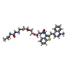  Mass: 608.791 Da / Num. of mol.: 1 / Source method: obtained synthetically / Formula: C33H44N4O5S Mass: 608.791 Da / Num. of mol.: 1 / Source method: obtained synthetically / Formula: C33H44N4O5S |
| #4: Chemical | ChemComp-K /   Mass: 39.098 Da / Num. of mol.: 1 / Source method: obtained synthetically / Formula: K Mass: 39.098 Da / Num. of mol.: 1 / Source method: obtained synthetically / Formula: K |
| Has ligand of interest | N |
-Experimental details
-Experiment
| Experiment | Method: X-RAY DIFFRACTION / Number of used crystals: 1 |
|---|
- Sample preparation
| Crystal | Density Matthews: 2.9 Å3/Da / Density % sol: 57.56 % |
|---|---|
| Crystal grow | Temperature: 294 K / Method: vapor diffusion, sitting drop / pH: 7.1 Details: 0.1 M Na cacodylate pH 7.1, 17.5% (w/v) polyethylene glycol (PEG) 3350, and 0.25 M Magnesium acetate |
-Data collection
| Diffraction | Mean temperature: 296 K / Ambient temp details: room temperature / Serial crystal experiment: Y |
|---|---|
| Diffraction source | Source: FREE ELECTRON LASER / Site: SLAC LCLS / Beamline: MFX / Wavelength: 1.303 Å |
| Detector | Type: DECTRIS EIGER X 16M / Detector: PIXEL / Date: Apr 8, 2018 |
| Radiation | Protocol: SINGLE WAVELENGTH / Monochromatic (M) / Laue (L): M / Scattering type: x-ray |
| Radiation wavelength | Wavelength: 1.303 Å / Relative weight: 1 |
| Reflection | Resolution: 3→52.5 Å / Num. obs: 5419 / % possible obs: 99.9 % / Redundancy: 3 % / Biso Wilson estimate: 56.8 Å2 / CC1/2: 0.7 / R split: 0.53 / Net I/σ(I): 2.3 |
| Reflection shell | Resolution: 3→3.3 Å / Redundancy: 3 % / Num. unique obs: 368 / CC1/2: 0.245 / R split: 1.421 / % possible all: 100 |
| Serial crystallography measurement | Focal spot size: 1 µm2 / Pulse duration: 0.044 fsec. |
| Serial crystallography sample delivery | Method: fixed target |
- Processing
| Software |
| |||||||||||||||||||||||||||||||||||
|---|---|---|---|---|---|---|---|---|---|---|---|---|---|---|---|---|---|---|---|---|---|---|---|---|---|---|---|---|---|---|---|---|---|---|---|---|
| Refinement | Method to determine structure: MOLECULAR REPLACEMENT Starting model: 6E8U Resolution: 3→51.64 Å / SU ML: 0.3945 / Cross valid method: FREE R-VALUE / σ(F): 1.34 / Phase error: 29.2294
| |||||||||||||||||||||||||||||||||||
| Solvent computation | Shrinkage radii: 0.9 Å / VDW probe radii: 1.11 Å | |||||||||||||||||||||||||||||||||||
| Displacement parameters | Biso mean: 45.19 Å2 | |||||||||||||||||||||||||||||||||||
| Refinement step | Cycle: LAST / Resolution: 3→51.64 Å
| |||||||||||||||||||||||||||||||||||
| Refine LS restraints |
| |||||||||||||||||||||||||||||||||||
| LS refinement shell |
|
