 Movie
Movie Controller

[English] 日本語
 Yorodumi
- PDB-6ppi: Kaposi's sarcoma-associated herpesvirus (KSHV), C12 portal dodeca... -
+ Open data

- Basic information
| Entry | Database: PDB / ID: 6ppi | |||||||||||||||||||||||||||||||||||||||||||||
|---|---|---|---|---|---|---|---|---|---|---|---|---|---|---|---|---|---|---|---|---|---|---|---|---|---|---|---|---|---|---|---|---|---|---|---|---|---|---|---|---|---|---|---|---|---|---|
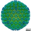
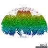
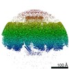
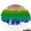
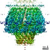
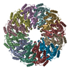
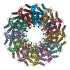
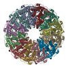
| Title | Kaposi's sarcoma-associated herpesvirus (KSHV), C12 portal dodecamer structure | |||||||||||||||||||||||||||||||||||||||||||||
 Components Components | Portal protein | |||||||||||||||||||||||||||||||||||||||||||||
 Keywords Keywords | VIRUS / portal / capsid / genome / genome packaging | |||||||||||||||||||||||||||||||||||||||||||||
| Function / homology | Herpesvirus portal protein / Herpesvirus UL6 like / chromosome organization / virion component / host cell nucleus / Portal protein / Core gene UL6 family protein Function and homology information Function and homology information | |||||||||||||||||||||||||||||||||||||||||||||
| Biological species |   Human herpesvirus 8 Human herpesvirus 8 | |||||||||||||||||||||||||||||||||||||||||||||
| Method | ELECTRON MICROSCOPY / single particle reconstruction / cryo EM / Resolution: 4.7 Å | |||||||||||||||||||||||||||||||||||||||||||||
 Authors Authors | Gong, D. / Dai, X. / Jih, J. / Liu, Y.T. / Bi, G.Q. / Sun, R. / Zhou, Z.H. | |||||||||||||||||||||||||||||||||||||||||||||
| Funding support |  United States, United States,  China, 14items China, 14items
| |||||||||||||||||||||||||||||||||||||||||||||
 Citation Citation | Journal: Cell / Year: 2019 Title: DNA-Packing Portal and Capsid-Associated Tegument Complexes in the Tumor Herpesvirus KSHV. Authors: Danyang Gong / Xinghong Dai / Jonathan Jih / Yun-Tao Liu / Guo-Qiang Bi / Ren Sun / Z Hong Zhou / Abstract: Assembly of Kaposi's sarcoma-associated herpesvirus (KSHV) begins at a bacteriophage-like portal complex that nucleates formation of an icosahedral capsid with capsid-associated tegument complexes ...Assembly of Kaposi's sarcoma-associated herpesvirus (KSHV) begins at a bacteriophage-like portal complex that nucleates formation of an icosahedral capsid with capsid-associated tegument complexes (CATCs) and facilitates translocation of an ∼150-kb dsDNA genome, followed by acquisition of a pleomorphic tegument and envelope. Because of deviation from icosahedral symmetry, KSHV portal and tegument structures have largely been obscured in previous studies. Using symmetry-relaxed cryo-EM, we determined the in situ structure of the KSHV portal and its interactions with surrounding capsid proteins, CATCs, and the terminal end of KSHV's dsDNA genome. Our atomic models of the portal and capsid/CATC, together with visualization of CATCs' variable occupancy and alternate orientation of CATC-interacting vertex triplexes, suggest a mechanism whereby the portal orchestrates procapsid formation and asymmetric long-range determination of CATC attachment during DNA packaging prior to pleomorphic tegumentation/envelopment. Structure-based mutageneses confirm that a triplex deep binding groove for CATCs is a hotspot that holds promise for antiviral development. | |||||||||||||||||||||||||||||||||||||||||||||
| History |
|
- Structure visualization
| Movie |
Movie viewer |
|---|---|
| Structure viewer | Molecule: MolmilJmol/JSmol |
- Downloads & links
-Download
| PDBx/mmCIF format | 6ppi.cif.gz | 881.7 KB | Display | PDBx/mmCIF format |
|---|---|---|---|---|
| PDB format | pdb6ppi.ent.gz | 717.4 KB | Display | PDB format |
| PDBx/mmJSON format | 6ppi.json.gz | Tree view | PDBx/mmJSON format | |
| Others |  Other downloads Other downloads |
-Validation report
| Arichive directory | https://data.pdbj.org/pub/pdb/validation_reports/pp/6ppiftp://data.pdbj.org/pub/pdb/validation_reports/pp/6ppi | HTTPS FTP |
|---|
-Related structure data
| Related structure data | 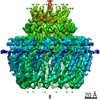 20437MC  6ppbC 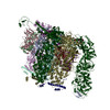 6ppdC 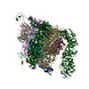 6pphC C: citing same article ( M: map data used to model this data |
|---|---|
| Similar structure data |


-Links
 PDBj
PDBj- Assembly
| Deposited unit | 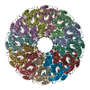
|
|---|---|
| 1 |
|
-Components
| #1: Protein | Mass: 68087.516 Da / Num. of mol.: 12 / Source method: isolated from a natural source Details: Subunit of portal complex present at KSHV's portal vertex; 12 copies constitute one dodecameric complex Source: (natural) Human herpesvirus 8 / Strain: GK18 / References: UniProt: Q76RH0, UniProt: F5HGK9*PLUS |
|---|
-Experimental details
-Experiment
| Experiment | Method: ELECTRON MICROSCOPY |
|---|---|
| EM experiment | Aggregation state: PARTICLE / 3D reconstruction method: single particle reconstruction |
- Sample preparation
| Component | Name: Human gammaherpesvirus 8 / Type: VIRUS / Entity ID: all / Source: NATURAL |
|---|---|
| Molecular weight | Experimental value: NO |
| Source (natural) | Organism: Human gammaherpesvirus 8 / Strain: BAC16 |
| Details of virus | Empty: NO / Enveloped: YES / Isolate: STRAIN / Type: VIRION |
| Natural host | Organism: Homo sapiens |
| Virus shell | Name: Capsid / Diameter: 1250 nm / Triangulation number (T number): 16 |
| Buffer solution | pH: 7.4 |
| Specimen | Embedding applied: NO / Shadowing applied: NO / Staining applied: NO / Vitrification applied: YES |
| Specimen support | Grid material: COPPER / Grid type: Quantifoil R2/1 |
| Vitrification | Instrument: HOMEMADE PLUNGER / Cryogen name: ETHANE / Chamber temperature: 298 K Details: The sample was manually blotted and frozen with a homemade plunger. |
- Electron microscopy imaging
| Experimental equipment |  Model: Titan Krios / Image courtesy: FEI Company |
|---|---|
| Microscopy | Model: FEI TITAN KRIOS |
| Electron gun | Electron source:  FIELD EMISSION GUN / Accelerating voltage: 300 kV / Illumination mode: FLOOD BEAM FIELD EMISSION GUN / Accelerating voltage: 300 kV / Illumination mode: FLOOD BEAM |
| Electron lens | Mode: BRIGHT FIELD / Nominal magnification: 14000 X / Calibrated magnification: 24271 X / Cs: 2.7 mm |
| Specimen holder | Cryogen: NITROGEN / Specimen holder model: FEI TITAN KRIOS AUTOGRID HOLDER / Temperature (max): 79 K |
| Image recording | Average exposure time: 13 sec. / Electron dose: 25 e/Å2 / Film or detector model: GATAN K2 SUMMIT (4k x 4k) / Num. of real images: 8007 |
| Image scans | Sampling size: 2.5 µm / Width: 1440 / Height: 1440 / Movie frames/image: 26 |
- Processing
| Software |
| ||||||||||||||||||||||||||||||||||||||||||||
|---|---|---|---|---|---|---|---|---|---|---|---|---|---|---|---|---|---|---|---|---|---|---|---|---|---|---|---|---|---|---|---|---|---|---|---|---|---|---|---|---|---|---|---|---|---|
| EM software |
| ||||||||||||||||||||||||||||||||||||||||||||
| CTF correction | Type: PHASE FLIPPING AND AMPLITUDE CORRECTION | ||||||||||||||||||||||||||||||||||||||||||||
| Particle selection | Num. of particles selected: 44328 | ||||||||||||||||||||||||||||||||||||||||||||
| Symmetry | Point symmetry: C12 (12 fold cyclic) | ||||||||||||||||||||||||||||||||||||||||||||
| 3D reconstruction | Resolution: 4.7 Å / Resolution method: FSC 0.143 CUT-OFF / Num. of particles: 39073 / Algorithm: FOURIER SPACE / Symmetry type: POINT | ||||||||||||||||||||||||||||||||||||||||||||
| Atomic model building | B value: 200 / Protocol: AB INITIO MODEL / Space: REAL / Target criteria: Correlation coefficient | ||||||||||||||||||||||||||||||||||||||||||||
| Refinement | Stereochemistry target values: GeoStd + Monomer Library | ||||||||||||||||||||||||||||||||||||||||||||
| Refine LS restraints |
|
