 Movie
Movie Controller


+ Open data

- Basic information
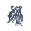
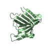
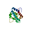
| Entry | Database: PDB / ID: 6pnw | |||||||||
|---|---|---|---|---|---|---|---|---|---|---|
| Title | X-RAY STRUCTURE OF ERABUTOXIN C, A DIMERIC NEUROTOXIN | |||||||||
 Components Components | Erabutoxin c | |||||||||
 Keywords Keywords | TOXIN / NEUROTOXIN / SNAKE VENOM | |||||||||
| Function / homology |  Function and homology information Function and homology informationacetylcholine receptor inhibitor activity / ion channel regulator activity / toxin activity / extracellular region Similarity search - Function | |||||||||
| Biological species |  Laticauda semifasciata (broad-banded blue sea krait) Laticauda semifasciata (broad-banded blue sea krait) | |||||||||
| Method |  X-RAY DIFFRACTION / MOLECULAR REPLACEMENT / Resolution: 2 Å X-RAY DIFFRACTION / MOLECULAR REPLACEMENT / Resolution: 2 Å | |||||||||
 Authors Authors | Corfield, P.W.R. / Low, B.W. | |||||||||
| Funding support |  United States, 2items United States, 2items
| |||||||||
 Citation Citation | Journal: AM.CRYST.ASSOC.,ABSTR.PAPERS (ANNUAL MEETING) / Year: 1992 Title: The Structure Of Erabutoxin C At 2.1A Resolution Authors: Corfield, P.W.R. / Low, B.W. Abstract: Scalar: PC53 #1: Journal: Acta Crystallogr.,Sect.B / Year: 1992Title: Structure determination of a dimeric form of erabutoxin-b, crystallized from a thiocyanate solution. Authors: Saludjian, P. / Prange, T. / Navaza, J. / Menez, R. / Guilloteau, J.P. / Ries-Kautt, M. / Ducruix, A. | |||||||||
| History |
| |||||||||
| Remark 700 | SHEET EACH OF THE TWO CRYSTALLOGRAPHICALLY INDEPENDENT MOLECULES HAS FIVE BETA STRANS A, B, C, D, E ...SHEET EACH OF THE TWO CRYSTALLOGRAPHICALLY INDEPENDENT MOLECULES HAS FIVE BETA STRANS A, B, C, D, E ARRANGED INTO TWO BETA SHEETS A,B AND C,D,E. THE TWO MOLECULES ARE LINKED VIA FOUR MAIN-CHAIN HYDROGEN BONDS BETWEEN LEU52, CYS54, AND GLU56 ON EACH MOLECULE, WITH A PSEUDO TWO-FOLD AXIS RELATING THE TWO MOLECULES PASSING BETWEEN THE TWO HYDROGEN BONDS LINING CYS54 ON EACH MOLECULE. THESE FOUR HYDROGEN BONDS LEAD TO AN EXTENDED SIX-CHAIN BETA SHEET IN THE DIMER. |
- Structure visualization
| Structure viewer | Molecule: MolmilJmol/JSmol |
|---|
- Downloads & links
-Download
| PDBx/mmCIF format | 6pnw.cif.gz | 41.5 KB | Display | PDBx/mmCIF format |
|---|---|---|---|---|
| PDB format | pdb6pnw.ent.gz | 27.6 KB | Display | PDB format |
| PDBx/mmJSON format | 6pnw.json.gz | Tree view | PDBx/mmJSON format | |
| Others |  Other downloads Other downloads |
-Validation report
| Arichive directory | https://data.pdbj.org/pub/pdb/validation_reports/pn/6pnwftp://data.pdbj.org/pub/pdb/validation_reports/pn/6pnw | HTTPS FTP |
|---|
-Related structure data
| Related structure data |  3ebxS S: Starting model for refinement |
|---|---|
| Similar structure data |


-Links
 PDBj
PDBj

- Assembly
| Deposited unit | 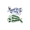
| ||||||||
|---|---|---|---|---|---|---|---|---|---|
| 1 |
| ||||||||
| Unit cell |
| ||||||||
| Noncrystallographic symmetry (NCS) | NCS oper: (Code: given / Matrix: (-1), |
-Components
| #1: Protein | Mass: 6862.682 Da / Num. of mol.: 2 / Source method: isolated from a natural source Source: (natural) Laticauda semifasciata (broad-banded blue sea krait)Secretion: VENOM / References: UniProt: Q7T2I5 #2: Water | ChemComp-HOH / |  Mass: 18.015 Da / Num. of mol.: 145 / Source method: isolated from a natural source / Formula: H2O Mass: 18.015 Da / Num. of mol.: 145 / Source method: isolated from a natural source / Formula: H2OHas protein modification | Y | |
|---|
-Experimental details
-Experiment
| Experiment | Method: X-RAY DIFFRACTION / Number of used crystals: 1 |
|---|
- Sample preparation
| Crystal | Density Matthews: 2.23 Å3/Da / Density % sol: 44.87 % Description: Long Rod, 0.25x0.35x1.5 mm; data collected on two halves |
|---|---|
| Crystal grow | Temperature: 298 K / Method: evaporation / pH: 6 / Details: PHOSPHATE BUFFER, AMMONIUM SULFATE PH 6 / PH range: 6 |
-Data collection
| Diffraction | Mean temperature: 290 K / Serial crystal experiment: N |
|---|---|
| Diffraction source | Source: SEALED TUBE / Type: OTHER / Details: Syntex P21 diffractometer / Wavelength: 1.542 Å |
| Detector | Type: SYNTEX / Detector: DIFFRACTOMETER / Date: Feb 1, 1983 / Details: Syntex P21 diffractometer |
| Radiation | Monochromator: GRAPHITE / Protocol: SINGLE WAVELENGTH / Monochromatic (M) / Laue (L): M / Scattering type: x-ray |
| Radiation wavelength | Wavelength: 1.542 Å / Relative weight: 1 |
| Reflection | Resolution: 2→38 Å / Num. obs: 7681 / % possible obs: 85.6 % / Redundancy: 2.2 % Details: Data processing used Bob Blessing's REFPK program for extracting intensities from the profiles, and his SORTAV for data merging. We did an empirical absorption correction based upon psi scans. Rmerge(I) obs: 0.08 / Net I/σ(I): 7.3 |
| Reflection shell | Resolution: 2→2.2 Å / Num. unique obs: 986 / % possible all: 70 |
- Processing
| Software |
| |||||||||||||||||||||
|---|---|---|---|---|---|---|---|---|---|---|---|---|---|---|---|---|---|---|---|---|---|---|
| Refinement | Method to determine structure: MOLECULAR REPLACEMENT Starting model: PDB entry 3EBX, ERABUTOXIN B Resolution: 2→10 Å / Cross valid method: NONE / σ(F): 4 Details: R Value from Prolsq based upon 5798 reflections with F>4Sigma. No Rfree set. The two crystallographically independent molecules are related by a non-crystallographic two-fold axis which ...Details: R Value from Prolsq based upon 5798 reflections with F>4Sigma. No Rfree set. The two crystallographically independent molecules are related by a non-crystallographic two-fold axis which lies in the YZ plane, at an approximate angle of 35 degrees with the Z axis. The mtrix transformations below willl operate on coordinates of atoms in chain B to give coordinates close to those for corresponding aotms in chain A. MTRIX1 1 -0.99795 0.02380 0.05933 -8.84136, MTRIX2 1 0.06391 0.35231 0.93370 8.38409 MTRIX3 1 0.00133 0.93558 -0.35311 -11.71019. Rotation and translational searches were done with Crowther and Blow's suite of programs applied locally.
| |||||||||||||||||||||
| Refinement step | Cycle: LAST / Resolution: 2→10 Å
|
