 Movie
Movie Controller

[English] 日本語
 Yorodumi
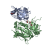
- PDB-6pnp: Crystal structure of the splice insert-free neurexin-1 LNS2 domai... -
+ Open data

- Basic information
| Entry | Database: PDB / ID: 6pnp | |||||||||
|---|---|---|---|---|---|---|---|---|---|---|
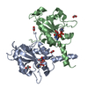
| Title | Crystal structure of the splice insert-free neurexin-1 LNS2 domain in complex with neurexophilin-1 | |||||||||
 Components Components |
| |||||||||
 Keywords Keywords | CELL ADHESION / Synapse / complex / splice-variant | |||||||||
| Function / homology |  Function and homology information Function and homology informationNeurexins and neuroligins / trans-synaptic protein complex / gephyrin clustering involved in postsynaptic density assembly / neuroligin clustering involved in postsynaptic membrane assembly / regulation of trans-synaptic signaling by endocannabinoid, modulating synaptic transmission / trans-synaptic signaling, modulating synaptic transmission / postsynaptic density protein 95 clustering / postsynaptic membrane assembly / neuroligin family protein binding / positive regulation of synapse maturation ...Neurexins and neuroligins / trans-synaptic protein complex / gephyrin clustering involved in postsynaptic density assembly / neuroligin clustering involved in postsynaptic membrane assembly / regulation of trans-synaptic signaling by endocannabinoid, modulating synaptic transmission / trans-synaptic signaling, modulating synaptic transmission / postsynaptic density protein 95 clustering / postsynaptic membrane assembly / neuroligin family protein binding / positive regulation of synapse maturation / regulation of grooming behavior / synaptic membrane adhesion / regulation of postsynaptic specialization assembly / presynapse assembly / regulation of insulin secretion involved in cellular response to glucose stimulus / regulation of postsynaptic density assembly / neurotransmitter secretion / acetylcholine receptor binding / vesicle docking involved in exocytosis / regulation of synaptic vesicle cycle / positive regulation of synapse assembly / adult behavior / neuromuscular process controlling balance / positive regulation of excitatory postsynaptic potential / regulation of presynapse assembly / synaptic cleft / prepulse inhibition / synapse assembly / cell adhesion molecule binding / presynaptic active zone membrane / calcium channel regulator activity / positive regulation of synaptic transmission, glutamatergic / learning / GABA-ergic synapse / cell projection / modulation of chemical synaptic transmission / Schaffer collateral - CA1 synapse / presynaptic membrane / chemical synaptic transmission / nuclear membrane / vesicle / signaling receptor binding / neuronal cell body / calcium ion binding / nucleolus / glutamatergic synapse / cell surface / endoplasmic reticulum / protein-containing complex / plasma membrane Similarity search - Function | |||||||||
| Biological species |  | |||||||||
| Method |  X-RAY DIFFRACTION / SYNCHROTRON / MOLECULAR REPLACEMENT / Resolution: 1.94 Å X-RAY DIFFRACTION / SYNCHROTRON / MOLECULAR REPLACEMENT / Resolution: 1.94 Å | |||||||||
 Authors Authors | Wilson, S.C. / White, K.I. / Zhou, Q. / Brunger, A.T. | |||||||||
| Funding support |  United States, 2items United States, 2items
| |||||||||
 Citation Citation | Journal: Embo J. / Year: 2019 Title: Structures of neurexophilin-neurexin complexes reveal a regulatory mechanism of alternative splicing. Authors: Wilson, S.C. / White, K.I. / Zhou, Q. / Pfuetzner, R.A. / Choi, U.B. / Sudhof, T.C. / Brunger, A.T. | |||||||||
| History |
|
- Structure visualization
| Structure viewer | Molecule: MolmilJmol/JSmol |
|---|
- Downloads & links
-Download
| PDBx/mmCIF format | 6pnp.cif.gz | 202 KB | Display | PDBx/mmCIF format |
|---|---|---|---|---|
| PDB format | pdb6pnp.ent.gz | 163.7 KB | Display | PDB format |
| PDBx/mmJSON format | 6pnp.json.gz | Tree view | PDBx/mmJSON format | |
| Others |  Other downloads Other downloads |
-Validation report
| Arichive directory | https://data.pdbj.org/pub/pdb/validation_reports/pn/6pnpftp://data.pdbj.org/pub/pdb/validation_reports/pn/6pnp | HTTPS FTP |
|---|
-Related structure data
| Related structure data |  6pnqC  2h0bS S: Starting model for refinement C: citing same article ( |
|---|---|
| Similar structure data |


-Links
 PDBj
PDBj


- Assembly
| Deposited unit | 
| ||||||||||
|---|---|---|---|---|---|---|---|---|---|---|---|
| 1 |
| ||||||||||
| Unit cell |
| ||||||||||
| Components on special symmetry positions |
|
-Components
| #1: Protein | Mass: 21133.787 Da / Num. of mol.: 1 / Mutation: C293A Source method: isolated from a genetically manipulated source Source: (gene. exp.)  Homo sapiens (human) / References: UniProt: Q9CS84 Homo sapiens (human) / References: UniProt: Q9CS84 |
|---|---|
| #2: Protein | Mass: 19081.398 Da / Num. of mol.: 1 / Mutation: N146D, N156D, N162D Source method: isolated from a genetically manipulated source Source: (gene. exp.) Homo sapiens (human) / References: UniProt: Q63366 |
| #3: Water | ChemComp-HOH /  Mass: 18.015 Da / Num. of mol.: 67 / Source method: isolated from a natural source / Formula: H2O Mass: 18.015 Da / Num. of mol.: 67 / Source method: isolated from a natural source / Formula: H2O |
| Has protein modification | Y |
-Experimental details
-Experiment
| Experiment | Method: X-RAY DIFFRACTION / Number of used crystals: 1 |
|---|
- Sample preparation
| Crystal | Density Matthews: 2.07 Å3/Da / Density % sol: 40.65 % |
|---|---|
| Crystal grow | Temperature: 298 K / Method: vapor diffusion, hanging drop / pH: 5 Details: 16% PEG 3350, 2% Tacsimate, and 0.1 M sodium citrate tribasic |
-Data collection
| Diffraction | Mean temperature: 100 K / Serial crystal experiment: N |
|---|---|
| Diffraction source | Source: SYNCHROTRON / Site: APS / Beamline: 24-ID-C / Wavelength: 0.9791 Å |
| Detector | Type: DECTRIS PILATUS 6M-F / Detector: PIXEL / Date: Mar 21, 2017 |
| Radiation | Protocol: SINGLE WAVELENGTH / Monochromatic (M) / Laue (L): M / Scattering type: x-ray |
| Radiation wavelength | Wavelength: 0.9791 Å / Relative weight: 1 |
| Reflection | Resolution: 1.94→45.6 Å / Num. obs: 24370 / % possible obs: 94.99 % / Redundancy: 13 % / Biso Wilson estimate: 39.5 Å2 / CC1/2: 0.999 / Rmerge(I) obs: 0.1 / Rpim(I) all: 0.02859 / Rrim(I) all: 0.1045 / Net I/σ(I): 14.05 |
| Reflection shell | Resolution: 1.94→2.01 Å / Redundancy: 9.1 % / Rmerge(I) obs: 1.214 / Mean I/σ(I) obs: 1.29 / Num. unique obs: 1662 / CC1/2: 0.796 / Rpim(I) all: 0.4089 / % possible all: 68.58 |
- Processing
| Software |
| ||||||||||||||||||||||||||||||||||||||||||||||||||||||||||||||||||||||||||||||||||||||||||
|---|---|---|---|---|---|---|---|---|---|---|---|---|---|---|---|---|---|---|---|---|---|---|---|---|---|---|---|---|---|---|---|---|---|---|---|---|---|---|---|---|---|---|---|---|---|---|---|---|---|---|---|---|---|---|---|---|---|---|---|---|---|---|---|---|---|---|---|---|---|---|---|---|---|---|---|---|---|---|---|---|---|---|---|---|---|---|---|---|---|---|---|
| Refinement | Method to determine structure: MOLECULAR REPLACEMENT Starting model: 2h0b Resolution: 1.94→45.6 Å / SU ML: 0.24 / Cross valid method: THROUGHOUT / σ(F): 1.36 / Phase error: 28.02
| ||||||||||||||||||||||||||||||||||||||||||||||||||||||||||||||||||||||||||||||||||||||||||
| Solvent computation | Shrinkage radii: 0.9 Å / VDW probe radii: 1.11 Å | ||||||||||||||||||||||||||||||||||||||||||||||||||||||||||||||||||||||||||||||||||||||||||
| Displacement parameters | Biso max: 315.29 Å2 / Biso mean: 76.06 Å2 / Biso min: 23.78 Å2 | ||||||||||||||||||||||||||||||||||||||||||||||||||||||||||||||||||||||||||||||||||||||||||
| Refinement step | Cycle: final / Resolution: 1.94→45.6 Å
| ||||||||||||||||||||||||||||||||||||||||||||||||||||||||||||||||||||||||||||||||||||||||||
| LS refinement shell | Refine-ID: X-RAY DIFFRACTION / Rfactor Rfree error: 0
| ||||||||||||||||||||||||||||||||||||||||||||||||||||||||||||||||||||||||||||||||||||||||||
| Refinement TLS params. | Method: refined / Refine-ID: X-RAY DIFFRACTION
| ||||||||||||||||||||||||||||||||||||||||||||||||||||||||||||||||||||||||||||||||||||||||||
| Refinement TLS group |
|
