 Movie
Movie Controller

[English] 日本語
 Yorodumi
- PDB-6pnl: Structure of Epimerase Mth375 from the thermophilic pseudomurein-... -
+ Open data

- Basic information
| Entry | Database: PDB / ID: 6pnl | ||||||
|---|---|---|---|---|---|---|---|
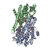
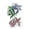
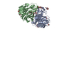
| Title | Structure of Epimerase Mth375 from the thermophilic pseudomurein-containing methanogen Methanothermobacter thermautotrophicus | ||||||
 Components Components | UDP-glucose 4-epimerase related protein | ||||||
 Keywords Keywords | ISOMERASE / Pseudomurein / UDP-N-acetylglucosamine / epimerase / cell wall / Methanothermobacter | ||||||
| Function / homology | NAD-dependent epimerase/dehydratase / NAD dependent epimerase/dehydratase family / NAD(P)-binding domain superfamily / nucleotide binding / NICOTINAMIDE-ADENINE-DINUCLEOTIDE / URIDINE-5'-DIPHOSPHATE / UDP-glucose 4-epimerase related protein Function and homology information Function and homology information | ||||||
| Biological species |   Methanothermobacter thermautotrophicus (archaea) Methanothermobacter thermautotrophicus (archaea) | ||||||
| Method |  X-RAY DIFFRACTION / SYNCHROTRON / MOLECULAR REPLACEMENT / molecular replacement / Resolution: 2.01 Å X-RAY DIFFRACTION / SYNCHROTRON / MOLECULAR REPLACEMENT / molecular replacement / Resolution: 2.01 Å | ||||||
 Authors Authors | Carbone, V. / Schofield, L.R. / Sutherland-Smith, A.J. / Ronimus, R.S. | ||||||
| Funding support |  New Zealand, 1items New Zealand, 1items
| ||||||
 Citation Citation | Journal: To Be Published Title: Structure of a UDP-GALE 4-epimerase (Mth375) from the thermophilic pseudomurein-containing methanogen Methanothermobacter thermautotrophicus Authors: Carbone, V. / Schofield, L.R. / Sutherland-Smith, A.J. / Ronimus, R.S. | ||||||
| History |
|
- Structure visualization
| Structure viewer | Molecule: MolmilJmol/JSmol |
|---|
- Downloads & links
-Download
| PDBx/mmCIF format | 6pnl.cif.gz | 95.3 KB | Display | PDBx/mmCIF format |
|---|---|---|---|---|
| PDB format | pdb6pnl.ent.gz | 68 KB | Display | PDB format |
| PDBx/mmJSON format | 6pnl.json.gz | Tree view | PDBx/mmJSON format | |
| Others |  Other downloads Other downloads |
-Validation report
| Summary document | 6pnl_validation.pdf.gz | 1.1 MB | Display | wwPDB validaton report |
|---|---|---|---|---|
| Full document | 6pnl_full_validation.pdf.gz | 1.1 MB | Display | |
| Data in XML | 6pnl_validation.xml.gz | 17.1 KB | Display | |
| Data in CIF | 6pnl_validation.cif.gz | 24.9 KB | Display | |
| Arichive directory | https://data.pdbj.org/pub/pdb/validation_reports/pn/6pnlftp://data.pdbj.org/pub/pdb/validation_reports/pn/6pnl | HTTPS FTP |
-Related structure data
| Related structure data | 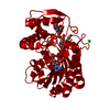 6pmhSC S: Starting model for refinement C: citing same article ( |
|---|---|
| Similar structure data |


-Links
 PDBj
PDBj


- Assembly
| Deposited unit | 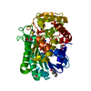
| ||||||||
|---|---|---|---|---|---|---|---|---|---|
| 1 | 
| ||||||||
| Unit cell |
| ||||||||
| Components on special symmetry positions |
|
-Components
-Protein , 1 types, 1 molecules A
| #1: Protein | Mass: 42049.320 Da / Num. of mol.: 1 Source method: isolated from a genetically manipulated source Source: (gene. exp.) Methanothermobacter thermautotrophicus (archaea)Production host:  |
|---|
-Non-polymers , 5 types, 245 molecules 


| #2: Chemical | ChemComp-NAD /  Mass: 663.425 Da / Num. of mol.: 1 / Source method: obtained synthetically / Formula: C21H27N7O14P2 / Feature type: SUBJECT OF INVESTIGATION / Comment: NAD*YM Mass: 663.425 Da / Num. of mol.: 1 / Source method: obtained synthetically / Formula: C21H27N7O14P2 / Feature type: SUBJECT OF INVESTIGATION / Comment: NAD*YM | ||||
|---|---|---|---|---|---|
| #3: Chemical | ChemComp-UDP /  Type: RNA linking / Mass: 404.161 Da / Num. of mol.: 1 / Source method: obtained synthetically / Formula: C9H14N2O12P2 / Feature type: SUBJECT OF INVESTIGATION / Comment: UDP*YM Type: RNA linking / Mass: 404.161 Da / Num. of mol.: 1 / Source method: obtained synthetically / Formula: C9H14N2O12P2 / Feature type: SUBJECT OF INVESTIGATION / Comment: UDP*YM | ||||
| #4: Chemical |  Mass: 62.068 Da / Num. of mol.: 3 / Source method: obtained synthetically / Formula: C2H6O2 Mass: 62.068 Da / Num. of mol.: 3 / Source method: obtained synthetically / Formula: C2H6O2#5: Chemical | ChemComp-SO4 / |  Mass: 96.063 Da / Num. of mol.: 1 / Source method: obtained synthetically / Formula: SO4 Mass: 96.063 Da / Num. of mol.: 1 / Source method: obtained synthetically / Formula: SO4#6: Water | ChemComp-HOH / | Mass: 18.015 Da / Num. of mol.: 239 / Source method: isolated from a natural source / Formula: H2O |
-Details
| Has ligand of interest | Y |
|---|---|
| Has protein modification | Y |
-Experimental details
-Experiment
| Experiment | Method: X-RAY DIFFRACTION / Number of used crystals: 1 |
|---|
- Sample preparation
| Crystal | Density Matthews: 2.4 Å3/Da / Density % sol: 48.78 % |
|---|---|
| Crystal grow | Temperature: 294 K / Method: vapor diffusion, sitting drop / pH: 7.5 Details: 0.1 M Sodium chloride, 1.6 M Ammonium sulfate 0.1 M Sodium HEPES, 3.0% w/v 1,8-Diaminooctane |
-Data collection
| Diffraction | Mean temperature: 100 K / Serial crystal experiment: N | ||||||||||||||||||||||||
|---|---|---|---|---|---|---|---|---|---|---|---|---|---|---|---|---|---|---|---|---|---|---|---|---|---|
| Diffraction source | Source: SYNCHROTRON / Site: Australian Synchrotron  / Beamline: MX2 / Wavelength: 0.953653 Å / Beamline: MX2 / Wavelength: 0.953653 Å | ||||||||||||||||||||||||
| Detector | Type: DECTRIS EIGER X 16M / Detector: PIXEL / Date: Feb 23, 2017 | ||||||||||||||||||||||||
| Radiation | Protocol: SINGLE WAVELENGTH / Monochromatic (M) / Laue (L): M / Scattering type: x-ray | ||||||||||||||||||||||||
| Radiation wavelength | Wavelength: 0.953653 Å / Relative weight: 1 | ||||||||||||||||||||||||
| Reflection | Resolution: 2.01→46.52 Å / Num. obs: 28060 / % possible obs: 99.7 % / Redundancy: 19.3 % / CC1/2: 0.996 / Rmerge(I) obs: 0.192 / Net I/σ(I): 16.1 | ||||||||||||||||||||||||
| Reflection shell | Diffraction-ID: 1
|
-Phasing
| Phasing | Method: molecular replacement | ||||||
|---|---|---|---|---|---|---|---|
| Phasing MR | R rigid body: 0.351
|
- Processing
| Software |
| ||||||||||||||||||||||||||||||||||||||||||||||||||||||||||||
|---|---|---|---|---|---|---|---|---|---|---|---|---|---|---|---|---|---|---|---|---|---|---|---|---|---|---|---|---|---|---|---|---|---|---|---|---|---|---|---|---|---|---|---|---|---|---|---|---|---|---|---|---|---|---|---|---|---|---|---|---|---|
| Refinement | Method to determine structure: MOLECULAR REPLACEMENT Starting model: 6PMH Resolution: 2.01→46.52 Å / Cor.coef. Fo:Fc: 0.959 / Cor.coef. Fo:Fc free: 0.944 / SU B: 3.015 / SU ML: 0.083 / SU R Cruickshank DPI: 0.1551 / Cross valid method: THROUGHOUT / σ(F): 0 / ESU R: 0.155 / ESU R Free: 0.133 Details: HYDROGENS HAVE BEEN ADDED IN THE RIDING POSITIONS U VALUES : REFINED INDIVIDUALLY
| ||||||||||||||||||||||||||||||||||||||||||||||||||||||||||||
| Solvent computation | Ion probe radii: 0.8 Å / Shrinkage radii: 0.8 Å / VDW probe radii: 1.2 Å | ||||||||||||||||||||||||||||||||||||||||||||||||||||||||||||
| Displacement parameters | Biso max: 68.01 Å2 / Biso mean: 22.501 Å2 / Biso min: 10.38 Å2
| ||||||||||||||||||||||||||||||||||||||||||||||||||||||||||||
| Refinement step | Cycle: final / Resolution: 2.01→46.52 Å
| ||||||||||||||||||||||||||||||||||||||||||||||||||||||||||||
| Refine LS restraints |
| ||||||||||||||||||||||||||||||||||||||||||||||||||||||||||||
| LS refinement shell | Resolution: 2.014→2.066 Å / Rfactor Rfree error: 0 / Total num. of bins used: 20
|
