 Movie
Movie Controller

[English] 日本語
 Yorodumi
- PDB-6pic: Crystal structure of Marinobacter subterrani acetylpolyamine amid... -
+ Open data

- Basic information
| Entry | Database: PDB / ID: 6pic | ||||||
|---|---|---|---|---|---|---|---|
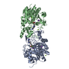
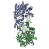
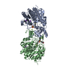
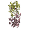
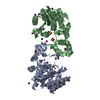
| Title | Crystal structure of Marinobacter subterrani acetylpolyamine amidohydrolase (msAPAH) complexed with 6-amino-N-hydroxyhexanamide | ||||||
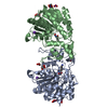
 Components Components | Acetylpolyamine Amidohydrolase | ||||||
 Keywords Keywords | HYDROLASE/INHIBITOR / acetylpolyamine amidohydrolase / polyamine deacetylase / hydrolase / hydrolase inhibitor / HYDROLASE-INHIBITOR complex | ||||||
| Function / homology |  Function and homology information Function and homology informationhistone deacetylase activity / epigenetic regulation of gene expression / hydrolase activity / metal ion binding Similarity search - Function | ||||||
| Biological species |  Marinobacter subterrani (bacteria) Marinobacter subterrani (bacteria) | ||||||
| Method |  X-RAY DIFFRACTION / SYNCHROTRON / MOLECULAR REPLACEMENT / Resolution: 2.031 Å X-RAY DIFFRACTION / SYNCHROTRON / MOLECULAR REPLACEMENT / Resolution: 2.031 Å | ||||||
 Authors Authors | Osko, J.D. / Christianson, D.W. | ||||||
| Funding support |  United States, 1items United States, 1items
| ||||||
 Citation Citation | Journal: Biochemistry / Year: 2019 Title: Structure and Function of the Acetylpolyamine Amidohydrolase from the Deep Earth HalophileMarinobacter subterrani. Authors: Osko, J.D. / Roose, B.W. / Shinsky, S.A. / Christianson, D.W. | ||||||
| History |
|
- Structure visualization
| Structure viewer | Molecule: MolmilJmol/JSmol |
|---|
- Downloads & links
-Download
| PDBx/mmCIF format | 6pic.cif.gz | 963.5 KB | Display | PDBx/mmCIF format |
|---|---|---|---|---|
| PDB format | pdb6pic.ent.gz | 793.8 KB | Display | PDB format |
| PDBx/mmJSON format | 6pic.json.gz | Tree view | PDBx/mmJSON format | |
| Others |  Other downloads Other downloads |
-Validation report
| Arichive directory | https://data.pdbj.org/pub/pdb/validation_reports/pi/6picftp://data.pdbj.org/pub/pdb/validation_reports/pi/6pic | HTTPS FTP |
|---|
-Related structure data
| Related structure data |  6phrC  6phtC  6phzC  6pi1C  6pi8C  6piaC 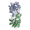 6pidC 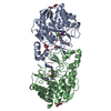 4zumS S: Starting model for refinement C: citing same article ( |
|---|---|
| Similar structure data |


-Links
 PDBj
PDBj- Assembly
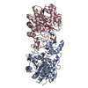
| Deposited unit | 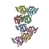
| ||||||||
|---|---|---|---|---|---|---|---|---|---|
| 1 | 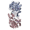
| ||||||||
| 2 | 
| ||||||||
| 3 | 
| ||||||||
| 4 | 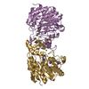
| ||||||||
| Unit cell |
|
-Components
-Protein , 1 types, 8 molecules ABCDEFGH
| #1: Protein | Mass: 37980.547 Da / Num. of mol.: 8 Source method: isolated from a genetically manipulated source Source: (gene. exp.) Marinobacter subterrani (bacteria) / Gene: Msub_13096 / Production host: |
|---|
-Non-polymers , 5 types, 684 molecules 


| #2: Chemical | ChemComp-ZN /  Mass: 65.409 Da / Num. of mol.: 8 / Source method: obtained synthetically / Formula: Zn Mass: 65.409 Da / Num. of mol.: 8 / Source method: obtained synthetically / Formula: Zn#3: Chemical | ChemComp-K /  Mass: 39.098 Da / Num. of mol.: 16 / Source method: obtained synthetically / Formula: K Mass: 39.098 Da / Num. of mol.: 16 / Source method: obtained synthetically / Formula: K#4: Chemical | ChemComp-MG /  Mass: 24.305 Da / Num. of mol.: 8 / Source method: obtained synthetically / Formula: Mg Mass: 24.305 Da / Num. of mol.: 8 / Source method: obtained synthetically / Formula: Mg#5: Chemical | ChemComp-6XA /  Mass: 146.188 Da / Num. of mol.: 8 / Source method: obtained synthetically / Formula: C6H14N2O2 / Feature type: SUBJECT OF INVESTIGATION Mass: 146.188 Da / Num. of mol.: 8 / Source method: obtained synthetically / Formula: C6H14N2O2 / Feature type: SUBJECT OF INVESTIGATION#6: Water | ChemComp-HOH / | Mass: 18.015 Da / Num. of mol.: 644 / Source method: isolated from a natural source / Formula: H2O |
|---|
-Details
| Has ligand of interest | Y |
|---|
-Experimental details
-Experiment
| Experiment | Method: X-RAY DIFFRACTION / Number of used crystals: 1 |
|---|
- Sample preparation
| Crystal | Density Matthews: 2.45 Å3/Da / Density % sol: 49.79 % / Description: plate-like crystals |
|---|---|
| Crystal grow | Temperature: 277 K / Method: vapor diffusion, sitting drop Details: 10 mg/ml msAPAH Protein, 0.2 M magnesium acetate tetrahydrate , 20% w/v PEG 3350, 1:1 ratio protein to precipitant |
-Data collection
| Diffraction | Mean temperature: 100 K / Serial crystal experiment: N |
|---|---|
| Diffraction source | Source: SYNCHROTRON / Site: SSRL / Beamline: BL12-2 / Wavelength: 0.98 Å |
| Detector | Type: DECTRIS PILATUS 6M / Detector: PIXEL / Date: Feb 22, 2019 |
| Radiation | Protocol: SINGLE WAVELENGTH / Monochromatic (M) / Laue (L): M / Scattering type: x-ray |
| Radiation wavelength | Wavelength: 0.98 Å / Relative weight: 1 |
| Reflection | Resolution: 2.03→66.97 Å / Num. obs: 154543 / % possible obs: 85.91 % / Redundancy: 1.9 % / Rpim(I) all: 0.158 / Net I/σ(I): 4.4 |
| Reflection shell | Resolution: 2.03→2.07 Å / Num. unique obs: 15944 / Rpim(I) all: 0.403 |
- Processing
| Software |
| |||||||||||||||||||||||||||||||||||||||||||||||||||||||||||||||||||||||||||||||||||||||||||||||||||||||||||||||||||||||||||||||||||||||||||||||||||||||||||||||||||||||||||||||||||||||||||||||||||||||||||||||||||||||||
|---|---|---|---|---|---|---|---|---|---|---|---|---|---|---|---|---|---|---|---|---|---|---|---|---|---|---|---|---|---|---|---|---|---|---|---|---|---|---|---|---|---|---|---|---|---|---|---|---|---|---|---|---|---|---|---|---|---|---|---|---|---|---|---|---|---|---|---|---|---|---|---|---|---|---|---|---|---|---|---|---|---|---|---|---|---|---|---|---|---|---|---|---|---|---|---|---|---|---|---|---|---|---|---|---|---|---|---|---|---|---|---|---|---|---|---|---|---|---|---|---|---|---|---|---|---|---|---|---|---|---|---|---|---|---|---|---|---|---|---|---|---|---|---|---|---|---|---|---|---|---|---|---|---|---|---|---|---|---|---|---|---|---|---|---|---|---|---|---|---|---|---|---|---|---|---|---|---|---|---|---|---|---|---|---|---|---|---|---|---|---|---|---|---|---|---|---|---|---|---|---|---|---|---|---|---|---|---|---|---|---|---|---|---|---|---|---|---|---|
| Refinement | Method to determine structure: MOLECULAR REPLACEMENT Starting model: PDB: 4ZUM Resolution: 2.031→51.973 Å / SU ML: 0.24 / Cross valid method: FREE R-VALUE / σ(F): 1.96 / Phase error: 24.51
| |||||||||||||||||||||||||||||||||||||||||||||||||||||||||||||||||||||||||||||||||||||||||||||||||||||||||||||||||||||||||||||||||||||||||||||||||||||||||||||||||||||||||||||||||||||||||||||||||||||||||||||||||||||||||
| Solvent computation | Shrinkage radii: 0.9 Å / VDW probe radii: 1.11 Å | |||||||||||||||||||||||||||||||||||||||||||||||||||||||||||||||||||||||||||||||||||||||||||||||||||||||||||||||||||||||||||||||||||||||||||||||||||||||||||||||||||||||||||||||||||||||||||||||||||||||||||||||||||||||||
| Refinement step | Cycle: LAST / Resolution: 2.031→51.973 Å
| |||||||||||||||||||||||||||||||||||||||||||||||||||||||||||||||||||||||||||||||||||||||||||||||||||||||||||||||||||||||||||||||||||||||||||||||||||||||||||||||||||||||||||||||||||||||||||||||||||||||||||||||||||||||||
| Refine LS restraints |
| |||||||||||||||||||||||||||||||||||||||||||||||||||||||||||||||||||||||||||||||||||||||||||||||||||||||||||||||||||||||||||||||||||||||||||||||||||||||||||||||||||||||||||||||||||||||||||||||||||||||||||||||||||||||||
| LS refinement shell |
| |||||||||||||||||||||||||||||||||||||||||||||||||||||||||||||||||||||||||||||||||||||||||||||||||||||||||||||||||||||||||||||||||||||||||||||||||||||||||||||||||||||||||||||||||||||||||||||||||||||||||||||||||||||||||
| Refinement TLS params. | Method: refined / Origin x: 55.138 Å / Origin y: -21.0913 Å / Origin z: 20.6044 Å
| |||||||||||||||||||||||||||||||||||||||||||||||||||||||||||||||||||||||||||||||||||||||||||||||||||||||||||||||||||||||||||||||||||||||||||||||||||||||||||||||||||||||||||||||||||||||||||||||||||||||||||||||||||||||||
| Refinement TLS group | Selection details: all |
