- PDB-6pf2: Crystal Structure of Amino Acids 1220-1276 of Human Beta Cardiac ... -
+
Open data
ID or keywords:
Loading...
-
Basic information
Entry
Database: PDB / ID: 6pf2
Title
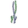
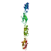
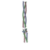
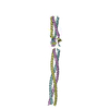
Crystal Structure of Amino Acids 1220-1276 of Human Beta Cardiac Myosin Fused to Gp7 and Eb1
Components
Myosin, heavy polypeptide 7, cardiac muscle, beta variant
Keywords
MOTOR PROTEIN / Myosin Rod / Myosin / Coiled-Coil / Gp7 / Eb1
Function / homology
Function and homology information
regulation of slow-twitch skeletal muscle fiber contraction / regulation of the force of skeletal muscle contraction / muscle myosin complex / regulation of the force of heart contraction / transition between fast and slow fiber / myosin filament / muscle filament sliding / cardiac muscle hypertrophy in response to stress / adult heart development / myosin complex ...regulation of slow-twitch skeletal muscle fiber contraction / regulation of the force of skeletal muscle contraction / muscle myosin complex / regulation of the force of heart contraction / transition between fast and slow fiber / myosin filament / muscle filament sliding / cardiac muscle hypertrophy in response to stress / adult heart development / myosin complex / myosin II complex / ventricular cardiac muscle tissue morphogenesis / microfilament motor activity / myofibril / ATP metabolic process / striated muscle contraction / skeletal muscle contraction / cardiac muscle contraction / stress fiber / regulation of heart rate / muscle contraction / sarcomere / Z disc / actin filament binding / calmodulin binding / ATP binding / cytoplasm Similarity search - Function
EB1-like C-terminal motif / DNA repair protein XRCC4-like, C-terminal / Myosin tail / Myosin tail / Myosin N-terminal SH3-like domain / Myosin S1 fragment, N-terminal / Myosin, N-terminal, SH3-like / Myosin N-terminal SH3-like domain profile. / Short calmodulin-binding motif containing conserved Ile and Gln residues. / IQ motif, EF-hand binding site ...EB1-like C-terminal motif / DNA repair protein XRCC4-like, C-terminal / Myosin tail / Myosin tail / Myosin N-terminal SH3-like domain / Myosin S1 fragment, N-terminal / Myosin, N-terminal, SH3-like / Myosin N-terminal SH3-like domain profile. / Short calmodulin-binding motif containing conserved Ile and Gln residues. / IQ motif, EF-hand binding site / Myosin motor domain profile. / Myosin head, motor domain / Myosin head (motor domain) / Myosin. Large ATPases. / IQ motif profile. / Kinesin motor domain superfamily / P-loop containing nucleoside triphosphate hydrolase Similarity search - Domain/homology
Mass: 18388.320 Da / Num. of mol.: 2 Source method: isolated from a genetically manipulated source Details: Human Beta cardiac myosin rod amino acids 1220-1276 with an amino-terminal fusion to Gp7 (amino acids 2-48, residue numbers 5-51) and carboxy-terminal fusion to Eb1 (amino acids 207-257, ...Details: Human Beta cardiac myosin rod amino acids 1220-1276 with an amino-terminal fusion to Gp7 (amino acids 2-48, residue numbers 5-51) and carboxy-terminal fusion to Eb1 (amino acids 207-257, residue numbers 2109-2159 ) Source: (gene. exp.) Homo sapiens (human) / Production host: Escherichia coli (E. coli) / References: UniProt: P12883*PLUS
Mass: 18.015 Da / Num. of mol.: 195 / Source method: isolated from a natural source / Formula: H2O
Has ligand of interest
N
Has protein modification
Y
-
Experimental details
-
Experiment
Experiment
Method: X-RAY DIFFRACTION / Number of used crystals: 1
-
Sample preparation
Crystal
Density Matthews: 3.03 Å3/Da / Density % sol: 59.43 %
Crystal grow
Temperature: 293 K / Method: vapor diffusion, hanging drop / pH: 8 Details: Crystals grew spontaneously at room temperature by mixing 1:1 ratio of 16 mg/ml protein in 100 mM NaCl, 10 mM HEPES pH 7.6, 0.1 mM TCEP with well solution containing 2.4 M ammonium phosphate ...Details: Crystals grew spontaneously at room temperature by mixing 1:1 ratio of 16 mg/ml protein in 100 mM NaCl, 10 mM HEPES pH 7.6, 0.1 mM TCEP with well solution containing 2.4 M ammonium phosphate pH 8.0, 100 mM HEPPS pH 8.0.
-
Data collection
Diffraction
Mean temperature: 100 K / Serial crystal experiment: N
In the structure databanks used in Yorodumi, some data are registered as the other names, "COVID-19 virus" and "2019-nCoV". Here are the details of the virus and the list of structure data.
Jan 31, 2019. EMDB accession codes are about to change! (news from PDBe EMDB page)
EMDB accession codes are about to change! (news from PDBe EMDB page)
The allocation of 4 digits for EMDB accession codes will soon come to an end. Whilst these codes will remain in use, new EMDB accession codes will include an additional digit and will expand incrementally as the available range of codes is exhausted. The current 4-digit format prefixed with “EMD-” (i.e. EMD-XXXX) will advance to a 5-digit format (i.e. EMD-XXXXX), and so on. It is currently estimated that the 4-digit codes will be depleted around Spring 2019, at which point the 5-digit format will come into force.
The EM Navigator/Yorodumi systems omit the EMD- prefix.
Related info.:Q: What is EMD? / ID/Accession-code notation in Yorodumi/EM Navigator
Yorodumi is a browser for structure data from EMDB, PDB, SASBDB, etc.
This page is also the successor to EM Navigator detail page, and also detail information page/front-end page for Omokage search.
The word "yorodu" (or yorozu) is an old Japanese word meaning "ten thousand". "mi" (miru) is to see.
Related info.:EMDB / PDB / SASBDB / Comparison of 3 databanks / Yorodumi Search / Aug 31, 2016. New EM Navigator & Yorodumi / Yorodumi Papers / Jmol/JSmol / Function and homology information / Changes in new EM Navigator and Yorodumi
 Movie
Movie Controller

 Yorodumi
 Open data
 Basic information
 Components
 Keywords
 Function and homology information
 Homo sapiens (human)
 X-RAY DIFFRACTION /
  Authors
 United States, 1items
  Citation
 Structure visualization
 Downloads & links
 Other downloads
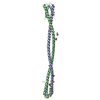


 PDBj
PDBj


 Assembly
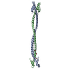


 Mass: 62.068 Da / Num. of mol.: 3 / Source method: obtained synthetically / Formula: C2H6O2
 Mass: 18.015 Da / Num. of mol.: 195 / Source method: isolated from a natural source / Formula: H2O
 Sample preparation
 Processing