モノクロメーター: DIAMOND / プロトコル: SINGLE WAVELENGTH / 単色(M)・ラウエ(L): M / 散乱光タイプ: x-ray
放射波長
波長: 0.9787 Å / 相対比: 1
反射
解像度: 2.051→39.742 Å / Num. obs: 16840 / % possible obs: 72.01 % / 冗長度: 8.68 % / Biso Wilson estimate: 45.815 Å2 詳細: Data were elliptically truncated with STARANISO. Statistics reported are for the observed reflections in spherical shells after apply the elliptical observation criterion. Elliptical ...詳細: Data were elliptically truncated with STARANISO. Statistics reported are for the observed reflections in spherical shells after apply the elliptical observation criterion. Elliptical completeness is 94% overall and 82% in the high resolution shell. CC1/2: 0.9963 / Rmerge(I) obs: 0.194 / Rrim(I) all: 0.207 / Χ2: 1.119 / Net I/σ(I): 9.29 / Num. measured all: 146211
解像度: 2.051→39.74 Å / SU ML: 0.21 / 交差検証法: THROUGHOUT / σ(F): 1.35 / 位相誤差: 23.36 / 立体化学のターゲット値: ML 詳細: 1. Data was aniostropically truncated with Staraniso. 2. Reference model restraints derrived from a higher resolution native structure, PDB-ID 2p02, were applied throughout. 3. While the ...詳細: 1. Data was aniostropically truncated with Staraniso. 2. Reference model restraints derrived from a higher resolution native structure, PDB-ID 2p02, were applied throughout. 3. While the crystallization drop was setup with protein in a buffer containing 0.002M ADP, the density in the ATP binding site had a break indicating that it was likely adenosine and not ADP bound. The density in the region of the phosphate binding site was disordered. In the end, it was modeled as a disordered pyrophosphate with a magnesium ion from the protein buffer and a potassium ion from the crystallization condition.
Rfactor
反射数
%反射
Selection details
Rfree
0.2029
825
4.9 %
RANDOM
Rwork
0.1726
16010
-
-
obs
0.1741
16835
72 %
-
溶媒の処理
減衰半径: 0.9 Å / VDWプローブ半径: 1.11 Å / 溶媒モデル: FLAT BULK SOLVENT MODEL
 ムービー
ムービー コントローラー


 データを開く
 基本情報
基本情報 要素
要素 キーワード
キーワード 機能・相同性情報
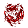
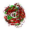
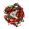
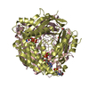
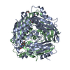
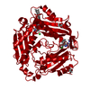
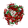
 Homo sapiens (ヒト)
 X線回折 /
  データ登録者
 米国, 3件
  引用
引用 構造の表示
構造の表示 ダウンロードとリンク
 その他のダウンロード
 PDBj
PDBj


 集合体
集合体


 分子量: 267.241 Da / 分子数: 1 / 由来タイプ: 合成 / 式: C10H13N5O4 / タイプ: SUBJECT OF INVESTIGATION
 分子量: 39.098 Da / 分子数: 1 / 由来タイプ: 合成 / 式: K
 分子量: 24.305 Da / 分子数: 1 / 由来タイプ: 合成 / 式: Mg / タイプ: SUBJECT OF INVESTIGATION
 分子量: 175.959 Da / 分子数: 1 / 由来タイプ: 合成 / 式: H2O7P2 / タイプ: SUBJECT OF INVESTIGATION
 試料調製
試料調製 解析
解析