 Movie
Movie Controller

[English] 日本語
 Yorodumi
- PDB-6p7y: Crystal Structure of the Cedar henipavirus Attachment G Glycoprot... -
+ Open data

- Basic information
| Entry | Database: PDB / ID: 6p7y | |||||||||
|---|---|---|---|---|---|---|---|---|---|---|
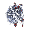
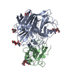
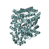
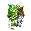
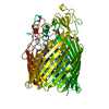
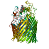
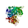
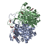
| Title | Crystal Structure of the Cedar henipavirus Attachment G Glycoprotein globular domain in complex with the receptor ephrin-B2 | |||||||||
 Components Components |
| |||||||||
 Keywords Keywords | VIRAL PROTEIN / Cedar virus / attachment / glycoprotein / G protein / receptor / ephrin-B2 / henipavirus | |||||||||
| Function / homology |  Function and homology information Function and homology informationpositive regulation of aorta morphogenesis / nephric duct morphogenesis / venous blood vessel morphogenesis / positive regulation of leukocyte adhesion to arterial endothelial cell / presynapse assembly / lymph vessel development / regulation of chemotaxis / positive regulation of cardiac muscle cell differentiation / cell migration involved in sprouting angiogenesis / adherens junction organization ...positive regulation of aorta morphogenesis / nephric duct morphogenesis / venous blood vessel morphogenesis / positive regulation of leukocyte adhesion to arterial endothelial cell / presynapse assembly / lymph vessel development / regulation of chemotaxis / positive regulation of cardiac muscle cell differentiation / cell migration involved in sprouting angiogenesis / adherens junction organization / blood vessel morphogenesis / EPH-Ephrin signaling / Ephrin signaling / regulation of postsynaptic neurotransmitter receptor internalization / exo-alpha-sialidase activity / keratinocyte proliferation / negative regulation of keratinocyte proliferation / EPH-ephrin mediated repulsion of cells / ephrin receptor signaling pathway / anatomical structure morphogenesis / regulation of postsynaptic membrane neurotransmitter receptor levels / ephrin receptor binding / T cell costimulation / EPHB-mediated forward signaling / axon guidance / animal organ morphogenesis / adherens junction / postsynaptic density membrane / Schaffer collateral - CA1 synapse / negative regulation of neuron projection development / cellular response to lipopolysaccharide / virus receptor activity / angiogenesis / presynaptic membrane / cell adhesion / host cell surface receptor binding / receptor ligand activity / focal adhesion / viral envelope / positive regulation of cell population proliferation / symbiont entry into host cell / dendrite / virion attachment to host cell / host cell plasma membrane / virion membrane / glutamatergic synapse / plasma membrane Similarity search - Function | |||||||||
| Biological species |  Cedar virus Cedar virus Homo sapiens (human) Homo sapiens (human) | |||||||||
| Method |  X-RAY DIFFRACTION / SYNCHROTRON / MOLECULAR REPLACEMENT / Resolution: 2.844 Å X-RAY DIFFRACTION / SYNCHROTRON / MOLECULAR REPLACEMENT / Resolution: 2.844 Å | |||||||||
 Authors Authors | Xu, K. / Nikolov, D.B. / Xu, Y. | |||||||||
 Citation Citation | Journal: Proc.Natl.Acad.Sci.USA / Year: 2019 Title: Structural and functional analyses reveal promiscuous and species specific use of ephrin receptors by Cedar virus. Authors: Laing, E.D. / Navaratnarajah, C.K. / Cheliout Da Silva, S. / Petzing, S.R. / Xu, Y. / Sterling, S.L. / Marsh, G.A. / Wang, L.F. / Amaya, M. / Nikolov, D.B. / Cattaneo, R. / Broder, C.C. / Xu, K. | |||||||||
| History |
|
- Structure visualization
| Structure viewer | Molecule: MolmilJmol/JSmol |
|---|
- Downloads & links
-Download
| PDBx/mmCIF format | 6p7y.cif.gz | 257.8 KB | Display | PDBx/mmCIF format |
|---|---|---|---|---|
| PDB format | pdb6p7y.ent.gz | 208.1 KB | Display | PDB format |
| PDBx/mmJSON format | 6p7y.json.gz | Tree view | PDBx/mmJSON format | |
| Others |  Other downloads Other downloads |
-Validation report
| Arichive directory | https://data.pdbj.org/pub/pdb/validation_reports/p7/6p7yftp://data.pdbj.org/pub/pdb/validation_reports/p7/6p7y | HTTPS FTP |
|---|
-Related structure data


-Links
 PDBj
PDBj


- Assembly
| Deposited unit | 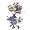
| ||||||||||||||||||||||||||||||||||||||||||||||||
|---|---|---|---|---|---|---|---|---|---|---|---|---|---|---|---|---|---|---|---|---|---|---|---|---|---|---|---|---|---|---|---|---|---|---|---|---|---|---|---|---|---|---|---|---|---|---|---|---|---|
| 1 | 
| ||||||||||||||||||||||||||||||||||||||||||||||||
| 2 | 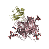
| ||||||||||||||||||||||||||||||||||||||||||||||||
| Unit cell |
| ||||||||||||||||||||||||||||||||||||||||||||||||
| Noncrystallographic symmetry (NCS) | NCS domain:
NCS domain segments:
NCS ensembles :
|
-Components
-Protein , 2 types, 4 molecules ACBD
| #1: Protein | Mass: 49042.242 Da / Num. of mol.: 2 / Fragment: globular domain (UNP residues 193-622) Source method: isolated from a genetically manipulated source Source: (gene. exp.) Cedar virusProduction host: Insect cell expression vector pTIE1 (others) References: UniProt: J7H333 #2: Protein | Mass: 16362.629 Da / Num. of mol.: 2 / Fragment: UNP residues 27-170 Source method: isolated from a genetically manipulated source Source: (gene. exp.) Homo sapiens (human) / Gene: EFNB2, EPLG5, HTKL, LERK5Production host: Insect cell expression vector pTIE1 (others) References: UniProt: P52799 |
|---|
-Sugars , 6 types, 13 molecules 
| #3: Polysaccharide | Source method: isolated from a genetically manipulated source #4: Polysaccharide | Source method: isolated from a genetically manipulated source #5: Polysaccharide | alpha-D-mannopyranose-(1-3)-[alpha-D-mannopyranose-(1-6)]beta-D-mannopyranose-(1-4)-2-acetamido-2- ...alpha-D-mannopyranose-(1-3)-[alpha-D-mannopyranose-(1-6)]beta-D-mannopyranose-(1-4)-2-acetamido-2-deoxy-beta-D-glucopyranose-(1-4)-[alpha-L-fucopyranose-(1-3)][alpha-L-fucopyranose-(1-6)]2-acetamido-2-deoxy-beta-D-glucopyranose | Source method: isolated from a genetically manipulated source #6: Polysaccharide | alpha-D-mannopyranose-(1-3)-[alpha-D-mannopyranose-(1-6)]beta-D-mannopyranose-(1-4)-2-acetamido-2- ...alpha-D-mannopyranose-(1-3)-[alpha-D-mannopyranose-(1-6)]beta-D-mannopyranose-(1-4)-2-acetamido-2-deoxy-beta-D-glucopyranose-(1-4)-2-acetamido-2-deoxy-beta-D-glucopyranose Source method: isolated from a genetically manipulated source #7: Polysaccharide | alpha-D-mannopyranose-(1-6)-beta-D-mannopyranose-(1-4)-2-acetamido-2-deoxy-beta-D-glucopyranose-(1- ...alpha-D-mannopyranose-(1-6)-beta-D-mannopyranose-(1-4)-2-acetamido-2-deoxy-beta-D-glucopyranose-(1-4)-2-acetamido-2-deoxy-beta-D-glucopyranose | Source method: isolated from a genetically manipulated source #8: Sugar |  Type: D-saccharide, beta linking / Mass: 221.208 Da / Num. of mol.: 2 Type: D-saccharide, beta linking / Mass: 221.208 Da / Num. of mol.: 2Source method: isolated from a genetically manipulated source Formula: C8H15NO6 |
|---|
-Non-polymers , 1 types, 161 molecules 
| #9: Water | ChemComp-HOH / Mass: 18.015 Da / Num. of mol.: 161 / Source method: isolated from a natural source / Formula: H2O |
|---|
-Details
| Has protein modification | Y |
|---|
-Experimental details
-Experiment
| Experiment | Method: X-RAY DIFFRACTION / Number of used crystals: 1 |
|---|
- Sample preparation
| Crystal | Density Matthews: 5.71 Å3/Da / Density % sol: 78.44 % |
|---|---|
| Crystal grow | Temperature: 298 K / Method: vapor diffusion, sitting drop / pH: 8 / Details: 100 mM Tris, pH 8.0, 18% PEG5000 MME |
-Data collection
| Diffraction | Mean temperature: 100 K / Serial crystal experiment: N | ||||||||||||||||||||||||||||||||||||||||||||||||||||||||||||||||||||||||||||||||||||||||
|---|---|---|---|---|---|---|---|---|---|---|---|---|---|---|---|---|---|---|---|---|---|---|---|---|---|---|---|---|---|---|---|---|---|---|---|---|---|---|---|---|---|---|---|---|---|---|---|---|---|---|---|---|---|---|---|---|---|---|---|---|---|---|---|---|---|---|---|---|---|---|---|---|---|---|---|---|---|---|---|---|---|---|---|---|---|---|---|---|---|
| Diffraction source | Source: SYNCHROTRON / Site: APS  / Beamline: 24-ID-C / Wavelength: 0.9192 Å / Beamline: 24-ID-C / Wavelength: 0.9192 Å | ||||||||||||||||||||||||||||||||||||||||||||||||||||||||||||||||||||||||||||||||||||||||
| Detector | Type: DECTRIS PILATUS 6M-F / Detector: PIXEL / Date: Jun 10, 2013 | ||||||||||||||||||||||||||||||||||||||||||||||||||||||||||||||||||||||||||||||||||||||||
| Radiation | Monochromator: double crystal Si(111) / Protocol: SINGLE WAVELENGTH / Monochromatic (M) / Laue (L): M / Scattering type: x-ray | ||||||||||||||||||||||||||||||||||||||||||||||||||||||||||||||||||||||||||||||||||||||||
| Radiation wavelength | Wavelength: 0.9192 Å / Relative weight: 1 | ||||||||||||||||||||||||||||||||||||||||||||||||||||||||||||||||||||||||||||||||||||||||
| Reflection | Resolution: 2.85→50 Å / Num. obs: 69486 / % possible obs: 100 % / Redundancy: 7.3 % / Rmerge(I) obs: 0.112 / Rpim(I) all: 0.053 / Rrim(I) all: 0.121 / Χ2: 1.36 / Net I/σ(I): 6.7 / Num. measured all: 505212 | ||||||||||||||||||||||||||||||||||||||||||||||||||||||||||||||||||||||||||||||||||||||||
| Reflection shell | Diffraction-ID: 1 / % possible all: 100
|
- Processing
| Software |
| |||||||||||||||||||||||||||||||||||||||||||||||||||||||||||||||||||||||||||||||||||||||||||||||||||||||||
|---|---|---|---|---|---|---|---|---|---|---|---|---|---|---|---|---|---|---|---|---|---|---|---|---|---|---|---|---|---|---|---|---|---|---|---|---|---|---|---|---|---|---|---|---|---|---|---|---|---|---|---|---|---|---|---|---|---|---|---|---|---|---|---|---|---|---|---|---|---|---|---|---|---|---|---|---|---|---|---|---|---|---|---|---|---|---|---|---|---|---|---|---|---|---|---|---|---|---|---|---|---|---|---|---|---|---|
| Refinement | Method to determine structure: MOLECULAR REPLACEMENT / Resolution: 2.844→45.973 Å / SU ML: 0.4 / Cross valid method: THROUGHOUT / σ(F): 1.35 / Phase error: 25.93
| |||||||||||||||||||||||||||||||||||||||||||||||||||||||||||||||||||||||||||||||||||||||||||||||||||||||||
| Solvent computation | Shrinkage radii: 0.9 Å / VDW probe radii: 1.11 Å | |||||||||||||||||||||||||||||||||||||||||||||||||||||||||||||||||||||||||||||||||||||||||||||||||||||||||
| Displacement parameters | Biso max: 152.45 Å2 / Biso mean: 65.29 Å2 / Biso min: 17.48 Å2 | |||||||||||||||||||||||||||||||||||||||||||||||||||||||||||||||||||||||||||||||||||||||||||||||||||||||||
| Refinement step | Cycle: final / Resolution: 2.844→45.973 Å
| |||||||||||||||||||||||||||||||||||||||||||||||||||||||||||||||||||||||||||||||||||||||||||||||||||||||||
| Refine LS restraints |
| |||||||||||||||||||||||||||||||||||||||||||||||||||||||||||||||||||||||||||||||||||||||||||||||||||||||||
| Refine LS restraints NCS |
| |||||||||||||||||||||||||||||||||||||||||||||||||||||||||||||||||||||||||||||||||||||||||||||||||||||||||
| LS refinement shell | Refine-ID: X-RAY DIFFRACTION / Rfactor Rfree error: 0 / Total num. of bins used: 14
|
