 Movie
Movie Controller

[English] 日本語
 Yorodumi
- PDB-6p2m: Crystal structure of the Caldicellulosiruptor lactoaceticus GH74 ... -
+ Open data

- Basic information
| Entry | Database: PDB / ID: 6p2m | |||||||||
|---|---|---|---|---|---|---|---|---|---|---|
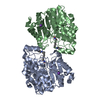
| Title | Crystal structure of the Caldicellulosiruptor lactoaceticus GH74 (ClGH74a) enzyme in complex with LLG xyloglucan | |||||||||
 Components Components | Type 3a cellulose-binding domain protein | |||||||||
 Keywords Keywords | HYDROLASE / glycosyl hydrolase / GH74 / xyloglucanase / 7-fold beta-propeller | |||||||||
| Function / homology |  Function and homology information Function and homology informationxyloglucan metabolic process / hydrolase activity, acting on glycosyl bonds / cellulose binding / polysaccharide catabolic process Similarity search - Function | |||||||||
| Biological species |  Caldicellulosiruptor lactoaceticus 6A (bacteria) Caldicellulosiruptor lactoaceticus 6A (bacteria) | |||||||||
| Method |  X-RAY DIFFRACTION / MOLECULAR REPLACEMENT / Resolution: 1.98 Å X-RAY DIFFRACTION / MOLECULAR REPLACEMENT / Resolution: 1.98 Å | |||||||||
 Authors Authors | Stogios, P.J. / Skarina, T. / Arnal, G. | |||||||||
| Funding support |  Canada, 1items Canada, 1items
| |||||||||
 Citation Citation | Journal: J.Biol.Chem. / Year: 2019 Title: Substrate specificity, regiospecificity, and processivity in glycoside hydrolase family 74. Authors: Arnal, G. / Stogios, P.J. / Asohan, J. / Attia, M.A. / Skarina, T. / Viborg, A.H. / Henrissat, B. / Savchenko, A. / Brumer, H. | |||||||||
| History |
|
- Structure visualization
| Structure viewer | Molecule: MolmilJmol/JSmol |
|---|
- Downloads & links
-Download
| PDBx/mmCIF format | 6p2m.cif.gz | 306.4 KB | Display | PDBx/mmCIF format |
|---|---|---|---|---|
| PDB format | pdb6p2m.ent.gz | 239.1 KB | Display | PDB format |
| PDBx/mmJSON format | 6p2m.json.gz | Tree view | PDBx/mmJSON format | |
| Others |  Other downloads Other downloads |
-Validation report
| Arichive directory | https://data.pdbj.org/pub/pdb/validation_reports/p2/6p2mftp://data.pdbj.org/pub/pdb/validation_reports/p2/6p2m | HTTPS FTP |
|---|
-Related structure data
| Related structure data |  6p2kC  6p2lC  6p2nC  6p2oC  5fkqS C: citing same article ( S: Starting model for refinement |
|---|---|
| Similar structure data |

-Links
 PDBj
PDBj
- Assembly
| Deposited unit | 
| ||||||||
|---|---|---|---|---|---|---|---|---|---|
| 1 |
| ||||||||
| Unit cell |
|
-Components
-Protein / Sugars , 2 types, 2 molecules A
| #1: Protein | Mass: 74671.812 Da / Num. of mol.: 1 Source method: isolated from a genetically manipulated source Source: (gene. exp.) Caldicellulosiruptor lactoaceticus 6A (bacteria)Gene: Calla_2311 / Plasmid: pMCSG53 / Production host: |
|---|---|
| #2: Polysaccharide | beta-D-galactopyranose-(1-2)-alpha-D-xylopyranose-(1-6)-beta-D-glucopyranose-(1-4)-[beta-D- ...beta-D-galactopyranose-(1-2)-alpha-D-xylopyranose-(1-6)-beta-D-glucopyranose-(1-4)-[beta-D-galactopyranose-(1-2)-alpha-D-xylopyranose-(1-6)]beta-D-glucopyranose-(1-4)-beta-D-glucopyranose Source method: isolated from a genetically manipulated source |
-Non-polymers , 4 types, 1186 molecules 


| #3: Chemical | ChemComp-PE3 /  Mass: 634.751 Da / Num. of mol.: 1 / Source method: obtained synthetically / Formula: C28H58O15 Mass: 634.751 Da / Num. of mol.: 1 / Source method: obtained synthetically / Formula: C28H58O15 | ||||
|---|---|---|---|---|---|
| #4: Chemical | ChemComp-GOL /  Mass: 92.094 Da / Num. of mol.: 4 / Source method: obtained synthetically / Formula: C3H8O3 Mass: 92.094 Da / Num. of mol.: 4 / Source method: obtained synthetically / Formula: C3H8O3#5: Chemical | ChemComp-TRS / |  Mass: 122.143 Da / Num. of mol.: 1 / Source method: obtained synthetically / Formula: C4H12NO3 / Comment: pH buffer*YM Mass: 122.143 Da / Num. of mol.: 1 / Source method: obtained synthetically / Formula: C4H12NO3 / Comment: pH buffer*YM#6: Water | ChemComp-HOH / | Mass: 18.015 Da / Num. of mol.: 1180 / Source method: isolated from a natural source / Formula: H2O |
-Experimental details
-Experiment
| Experiment | Method: X-RAY DIFFRACTION / Number of used crystals: 1 |
|---|
- Sample preparation
| Crystal | Density Matthews: 2.39 Å3/Da / Density % sol: 48.45 % |
|---|---|
| Crystal grow | Temperature: 298 K / Method: vapor diffusion, sitting drop / pH: 8.5 Details: 25% (w/v) PEG3350, 0.1 M Tris pH 8.5, xyloglucan mixture |
-Data collection
| Diffraction | Mean temperature: 100 K / Serial crystal experiment: N |
|---|---|
| Diffraction source | Source: ROTATING ANODE / Type: RIGAKU MICROMAX-007 / Wavelength: 1.5418 Å |
| Detector | Type: RIGAKU RAXIS IV / Detector: IMAGE PLATE / Date: Jul 20, 2016 |
| Radiation | Protocol: SINGLE WAVELENGTH / Monochromatic (M) / Laue (L): M / Scattering type: x-ray |
| Radiation wavelength | Wavelength: 1.5418 Å / Relative weight: 1 |
| Reflection | Resolution: 1.98→25 Å / Num. obs: 50820 / % possible obs: 100 % / Redundancy: 7.8 % / Rmerge(I) obs: 0.165 / Rpim(I) all: 0.063 / Net I/σ(I): 18.2 |
| Reflection shell | Resolution: 1.98→2.01 Å / Redundancy: 6.3 % / Rmerge(I) obs: 0.873 / Mean I/σ(I) obs: 2.4 / Num. unique obs: 2479 / CC1/2: 0.623 / Rpim(I) all: 0.438 / % possible all: 99.9 |
- Processing
| Software |
| ||||||||||||||||||||||||||||||||||||||||||||||||||||||||||||||||||||||||||||||||||||||||||||||||||||
|---|---|---|---|---|---|---|---|---|---|---|---|---|---|---|---|---|---|---|---|---|---|---|---|---|---|---|---|---|---|---|---|---|---|---|---|---|---|---|---|---|---|---|---|---|---|---|---|---|---|---|---|---|---|---|---|---|---|---|---|---|---|---|---|---|---|---|---|---|---|---|---|---|---|---|---|---|---|---|---|---|---|---|---|---|---|---|---|---|---|---|---|---|---|---|---|---|---|---|---|---|---|
| Refinement | Method to determine structure: MOLECULAR REPLACEMENT Starting model: 5fkq Resolution: 1.98→24.608 Å / SU ML: 0.19 / Cross valid method: FREE R-VALUE / σ(F): 0 / Phase error: 19.36
| ||||||||||||||||||||||||||||||||||||||||||||||||||||||||||||||||||||||||||||||||||||||||||||||||||||
| Solvent computation | Shrinkage radii: 0.9 Å / VDW probe radii: 1.11 Å | ||||||||||||||||||||||||||||||||||||||||||||||||||||||||||||||||||||||||||||||||||||||||||||||||||||
| Refinement step | Cycle: LAST / Resolution: 1.98→24.608 Å
| ||||||||||||||||||||||||||||||||||||||||||||||||||||||||||||||||||||||||||||||||||||||||||||||||||||
| Refine LS restraints |
| ||||||||||||||||||||||||||||||||||||||||||||||||||||||||||||||||||||||||||||||||||||||||||||||||||||
| LS refinement shell |
| ||||||||||||||||||||||||||||||||||||||||||||||||||||||||||||||||||||||||||||||||||||||||||||||||||||
| Refinement TLS params. | Method: refined / Refine-ID: X-RAY DIFFRACTION
| ||||||||||||||||||||||||||||||||||||||||||||||||||||||||||||||||||||||||||||||||||||||||||||||||||||
| Refinement TLS group |
|
