 Movie
Movie Controller

[English] 日本語
 Yorodumi
- PDB-6p13: Structure of spastin AAA domain (T692A mutant) in complex with a ... -
+ Open data

- Basic information
| Entry | Database: PDB / ID: 6p13 | |||||||||
|---|---|---|---|---|---|---|---|---|---|---|
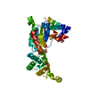
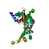
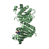
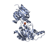
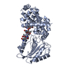
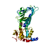
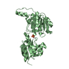
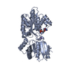
| Title | Structure of spastin AAA domain (T692A mutant) in complex with a diaminotriazole-based inhibitor (crystal form A) | |||||||||
 Components Components | Spastin | |||||||||
 Keywords Keywords | ISOMERASE/ISOMERASE INHIBITOR / inhibitor / complex / AAA protein / ISOMERASE-ISOMERASE INHIBITOR complex | |||||||||
| Function / homology |  Function and homology information Function and homology informationhemocyte migration / positive regulation of axon extension involved in regeneration / Sealing of the nuclear envelope (NE) by ESCRT-III / negative regulation of synaptic assembly at neuromuscular junction / negative regulation of neuromuscular synaptic transmission / positive regulation of neuromuscular synaptic transmission / microtubule-severing ATPase / positive regulation of synaptic assembly at neuromuscular junction / microtubule severing ATPase activity / regulation of terminal button organization ...hemocyte migration / positive regulation of axon extension involved in regeneration / Sealing of the nuclear envelope (NE) by ESCRT-III / negative regulation of synaptic assembly at neuromuscular junction / negative regulation of neuromuscular synaptic transmission / positive regulation of neuromuscular synaptic transmission / microtubule-severing ATPase / positive regulation of synaptic assembly at neuromuscular junction / microtubule severing ATPase activity / regulation of terminal button organization / central nervous system neuron axonogenesis / microtubule severing / positive regulation of microtubule depolymerization / mitotic chromosome movement towards spindle pole / mitotic spindle elongation / negative regulation of microtubule depolymerization / positive regulation of dendrite morphogenesis / protein hexamerization / positive regulation of lipid metabolic process / mitotic sister chromatid segregation / alpha-tubulin binding / adult locomotory behavior / lipid droplet / neuromuscular junction / locomotory behavior / microtubule cytoskeleton organization / spindle / terminal bouton / microtubule cytoskeleton / chromosome / microtubule binding / microtubule / cell division / centrosome / ATP hydrolysis activity / ATP binding / membrane / cytoplasm Similarity search - Function | |||||||||
| Biological species |  | |||||||||
| Method |  X-RAY DIFFRACTION / SYNCHROTRON / MOLECULAR REPLACEMENT / Resolution: 2.1 Å X-RAY DIFFRACTION / SYNCHROTRON / MOLECULAR REPLACEMENT / Resolution: 2.1 Å | |||||||||
 Authors Authors | Pisa, R. / Cupido, T. / Kapoor, T.M. | |||||||||
| Funding support |  United States, 2items United States, 2items
| |||||||||
 Citation Citation | Journal: Cell Chem Biol / Year: 2019 Title: Analyzing Resistance to Design Selective Chemical Inhibitors for AAA Proteins. Authors: Pisa, R. / Cupido, T. / Steinman, J.B. / Jones, N.H. / Kapoor, T.M. | |||||||||
| History |
|
- Structure visualization
| Structure viewer | Molecule: MolmilJmol/JSmol |
|---|
- Downloads & links
-Download
| PDBx/mmCIF format | 6p13.cif.gz | 74.9 KB | Display | PDBx/mmCIF format |
|---|---|---|---|---|
| PDB format | pdb6p13.ent.gz | 51.5 KB | Display | PDB format |
| PDBx/mmJSON format | 6p13.json.gz | Tree view | PDBx/mmJSON format | |
| Others |  Other downloads Other downloads |
-Validation report
| Arichive directory | https://data.pdbj.org/pub/pdb/validation_reports/p1/6p13ftp://data.pdbj.org/pub/pdb/validation_reports/p1/6p13 | HTTPS FTP |
|---|
-Related structure data
| Related structure data |  6p10C  6p11C  6p12C  6p14C  3b9pS S: Starting model for refinement C: citing same article ( |
|---|---|
| Similar structure data |


-Links
 PDBj
PDBj


- Assembly
| Deposited unit | 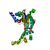
| ||||||||
|---|---|---|---|---|---|---|---|---|---|
| 1 |
| ||||||||
| Unit cell |
|
-Components
| #1: Protein | Mass: 34291.004 Da / Num. of mol.: 1 / Fragment: AAA domain (UNP residues 445-758) / Mutation: T692A Source method: isolated from a genetically manipulated source Source: (gene. exp.)  |
|---|---|
| #2: Chemical | ChemComp-SO4 /   Mass: 96.063 Da / Num. of mol.: 1 / Source method: obtained synthetically / Formula: SO4 Mass: 96.063 Da / Num. of mol.: 1 / Source method: obtained synthetically / Formula: SO4 |
| #3: Chemical | ChemComp-NKY /   Mass: 384.364 Da / Num. of mol.: 1 / Source method: obtained synthetically / Formula: C18H17FN6O3 / Feature type: SUBJECT OF INVESTIGATION Mass: 384.364 Da / Num. of mol.: 1 / Source method: obtained synthetically / Formula: C18H17FN6O3 / Feature type: SUBJECT OF INVESTIGATION |
| #4: Chemical | ChemComp-MPD / (  Mass: 118.174 Da / Num. of mol.: 1 / Source method: obtained synthetically / Formula: C6H14O2 / Comment: precipitant*YM Mass: 118.174 Da / Num. of mol.: 1 / Source method: obtained synthetically / Formula: C6H14O2 / Comment: precipitant*YM |
| #5: Water | ChemComp-HOH /  Mass: 18.015 Da / Num. of mol.: 108 / Source method: isolated from a natural source / Formula: H2O Mass: 18.015 Da / Num. of mol.: 108 / Source method: isolated from a natural source / Formula: H2O |
| Has ligand of interest | Y |
-Experimental details
-Experiment
| Experiment | Method: X-RAY DIFFRACTION / Number of used crystals: 1 |
|---|
- Sample preparation
| Crystal | Density Matthews: 2.58 Å3/Da / Density % sol: 52.26 % |
|---|---|
| Crystal grow | Temperature: 291 K / Method: vapor diffusion, hanging drop Details: 0.1 M sodium acetate, pH 5.0-6.5, 2% PEG4000, 15% MPD PH range: 5.0-6.5 |
-Data collection
| Diffraction | Mean temperature: 100 K / Serial crystal experiment: N |
|---|---|
| Diffraction source | Source: SYNCHROTRON / Site: NSLS-II / Beamline: 17-ID-1 / Wavelength: 0.9201 Å |
| Detector | Type: DECTRIS EIGER X 9M / Detector: PIXEL / Date: Jun 28, 2018 |
| Diffraction measurement | Details: 1.00 degrees, 0.20 sec, detector distance 220.00 mm Method: \w scans |
| Radiation | Protocol: SINGLE WAVELENGTH / Monochromatic (M) / Laue (L): M / Scattering type: x-ray |
| Radiation wavelength | Wavelength: 0.9201 Å / Relative weight: 1 |
| Reflection | Av R equivalents: 0.052 / Number: 400597 |
| Reflection | Resolution: 2.1→50 Å / Num. obs: 20423 / % possible obs: 99.9 % / Observed criterion σ(F): 0 / Redundancy: 5.8 % / Biso Wilson estimate: 46.0316109984 Å2 / Rmerge(I) obs: 0.043 / Net I/av σ(I): 42 / Net I/σ(I): 13.4 |
| Reflection shell | Resolution: 2.1→2.14 Å / Redundancy: 6 % / Rmerge(I) obs: 0.448 / Mean I/σ(I) obs: 2.7 / Num. unique obs: 1015 / CC1/2: 0.869 / % possible all: 100 |
| Cell measurement | Reflection used: 400597 |
- Processing
| Software |
| ||||||||||||||||||||||||||||||||||||||||||||||||||||||||||||||||||||||||||||||||||||||||||||||||
|---|---|---|---|---|---|---|---|---|---|---|---|---|---|---|---|---|---|---|---|---|---|---|---|---|---|---|---|---|---|---|---|---|---|---|---|---|---|---|---|---|---|---|---|---|---|---|---|---|---|---|---|---|---|---|---|---|---|---|---|---|---|---|---|---|---|---|---|---|---|---|---|---|---|---|---|---|---|---|---|---|---|---|---|---|---|---|---|---|---|---|---|---|---|---|---|---|---|
| Refinement | Method to determine structure: MOLECULAR REPLACEMENT Starting model: PDB entry 3B9P Resolution: 2.1→39.849 Å / SU ML: 0.27 / Cross valid method: THROUGHOUT / σ(F): 1.38 / Phase error: 27.06
| ||||||||||||||||||||||||||||||||||||||||||||||||||||||||||||||||||||||||||||||||||||||||||||||||
| Solvent computation | Shrinkage radii: 0.9 Å / VDW probe radii: 1.11 Å | ||||||||||||||||||||||||||||||||||||||||||||||||||||||||||||||||||||||||||||||||||||||||||||||||
| Displacement parameters | Biso max: 93.79 Å2 / Biso mean: 50.7305 Å2 / Biso min: 27.48 Å2 | ||||||||||||||||||||||||||||||||||||||||||||||||||||||||||||||||||||||||||||||||||||||||||||||||
| Refinement step | Cycle: final / Resolution: 2.1→39.849 Å
| ||||||||||||||||||||||||||||||||||||||||||||||||||||||||||||||||||||||||||||||||||||||||||||||||
| Refine LS restraints |
| ||||||||||||||||||||||||||||||||||||||||||||||||||||||||||||||||||||||||||||||||||||||||||||||||
| LS refinement shell | Refine-ID: X-RAY DIFFRACTION / Rfactor Rfree error: 0
|
