 Movie
Movie Controller

[English] 日本語
 Yorodumi
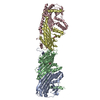
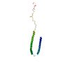
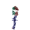
- PDB-6ov6: CRYSTALLOGRAPHIC STRUCTURE OF THE C24 PROTEIN FROM THE ANTARCTIC ... -
+ Open data

- Basic information
| Entry | Database: PDB / ID: 6ov6 | ||||||
|---|---|---|---|---|---|---|---|
| Title | CRYSTALLOGRAPHIC STRUCTURE OF THE C24 PROTEIN FROM THE ANTARCTIC MICROORGANISM BIZIONIA ARGENTINENSIS | ||||||
 Components Components | C24 PROTEIN | ||||||
 Keywords Keywords | UNKNOWN FUNCTION / FIBER / VIRAL ORIGIN | ||||||
| Function / homology | metal ion binding / : / Tail fiber protein Function and homology information Function and homology information | ||||||
| Biological species |  Bizionia argentinensis JUB59 (bacteria) Bizionia argentinensis JUB59 (bacteria) | ||||||
| Method |  X-RAY DIFFRACTION / SYNCHROTRON / SAD / Resolution: 1.82 Å X-RAY DIFFRACTION / SYNCHROTRON / SAD / Resolution: 1.82 Å | ||||||
 Authors Authors | Klinke, S. / Rinaldi, J. / Guimaraes, B.G. / Pellizza, L. / Aran, M. | ||||||
 Citation Citation | Journal: J.Struct.Biol. / Year: 2020 Title: Structure of the putative long tail fiber receptor-binding tip of a novel temperate bacteriophage from the Antarctic bacterium Bizionia argentinensis JUB59. Authors: Pellizza, L. / Lopez, J.L. / Vazquez, S. / Sycz, G. / Guimaraes, B.G. / Rinaldi, J. / Goldbaum, F.A. / Aran, M. / Mac Cormack, W.P. / Klinke, S. | ||||||
| History |
|
- Structure visualization
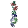
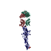
| Structure viewer | Molecule: MolmilJmol/JSmol |
|---|
- Downloads & links
-Download
| PDBx/mmCIF format | 6ov6.cif.gz | 164.1 KB | Display | PDBx/mmCIF format |
|---|---|---|---|---|
| PDB format | pdb6ov6.ent.gz | 128.1 KB | Display | PDB format |
| PDBx/mmJSON format | 6ov6.json.gz | Tree view | PDBx/mmJSON format | |
| Others |  Other downloads Other downloads |
-Validation report
| Arichive directory | https://data.pdbj.org/pub/pdb/validation_reports/ov/6ov6ftp://data.pdbj.org/pub/pdb/validation_reports/ov/6ov6 | HTTPS FTP |
|---|
-Related structure data
| Similar structure data |
|---|
-Links
 PDBj
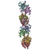
PDBj- Assembly
| Deposited unit | 
| ||||||||
|---|---|---|---|---|---|---|---|---|---|
| 1 |
| ||||||||
| Unit cell |
|
-Components
| #1: Protein | Mass: 29729.789 Da / Num. of mol.: 3 Source method: isolated from a genetically manipulated source Source: (gene. exp.) Bizionia argentinensis JUB59 (bacteria)Gene: BZARG_797 / Plasmid: PDEST-527 / Production host: #2: Chemical | ChemComp-MN / |   Mass: 54.938 Da / Num. of mol.: 1 / Source method: obtained synthetically / Formula: Mn Mass: 54.938 Da / Num. of mol.: 1 / Source method: obtained synthetically / Formula: Mn#3: Water | ChemComp-HOH / |  Mass: 18.015 Da / Num. of mol.: 600 / Source method: isolated from a natural source / Formula: H2O Mass: 18.015 Da / Num. of mol.: 600 / Source method: isolated from a natural source / Formula: H2O |
|---|
-Experimental details
-Experiment
| Experiment | Method: X-RAY DIFFRACTION / Number of used crystals: 1 |
|---|
- Sample preparation
| Crystal | Density Matthews: 2.77 Å3/Da / Density % sol: 55.53 % / Description: Rhomboidal-shaped crystals |
|---|---|
| Crystal grow | Temperature: 295 K / Method: vapor diffusion, hanging drop / pH: 7.5 Details: 12% PEG 8000, 0.1 M SODIUM CITRATE, 0.1 M AMMONIUM SULFATE PH range: 7.5 |
-Data collection
| Diffraction |
| ||||||||||||||||||
|---|---|---|---|---|---|---|---|---|---|---|---|---|---|---|---|---|---|---|---|
| Diffraction source |
| ||||||||||||||||||
| Detector |
| ||||||||||||||||||
| Radiation |
| ||||||||||||||||||
| Radiation wavelength |
| ||||||||||||||||||
| Reflection | Resolution: 1.82→50 Å / Num. obs: 87380 / % possible obs: 97.2 % / Observed criterion σ(I): 0 / Redundancy: 5.6 % / Biso Wilson estimate: 33.28 Å2 / Rmerge(I) obs: 0.053 / Rsym value: 0.053 / Net I/σ(I): 17.3 | ||||||||||||||||||
| Reflection shell | Resolution: 1.82→1.93 Å / Redundancy: 5.6 % / Rmerge(I) obs: 0.593 / Mean I/σ(I) obs: 2.4 / Num. unique obs: 6246 / Rsym value: 0.593 / % possible all: 95.6 |

- Processing
| Software |
| ||||||||||||||||||||||||||||||||||||||||||||||||||||||||||||||||||||||||||||||||||||||||||||||||||||||||||||||||||
|---|---|---|---|---|---|---|---|---|---|---|---|---|---|---|---|---|---|---|---|---|---|---|---|---|---|---|---|---|---|---|---|---|---|---|---|---|---|---|---|---|---|---|---|---|---|---|---|---|---|---|---|---|---|---|---|---|---|---|---|---|---|---|---|---|---|---|---|---|---|---|---|---|---|---|---|---|---|---|---|---|---|---|---|---|---|---|---|---|---|---|---|---|---|---|---|---|---|---|---|---|---|---|---|---|---|---|---|---|---|---|---|---|---|---|---|
| Refinement | Method to determine structure: SAD / Resolution: 1.82→44.12 Å / Cor.coef. Fo:Fc: 0.952 / Cor.coef. Fo:Fc free: 0.937 / SU R Cruickshank DPI: 0.11 / Cross valid method: THROUGHOUT / σ(F): 0
| ||||||||||||||||||||||||||||||||||||||||||||||||||||||||||||||||||||||||||||||||||||||||||||||||||||||||||||||||||
| Displacement parameters | Biso mean: 38.1 Å2
| ||||||||||||||||||||||||||||||||||||||||||||||||||||||||||||||||||||||||||||||||||||||||||||||||||||||||||||||||||
| Refine analyze | Luzzati coordinate error obs: 0.24 Å | ||||||||||||||||||||||||||||||||||||||||||||||||||||||||||||||||||||||||||||||||||||||||||||||||||||||||||||||||||
| Refinement step | Cycle: LAST / Resolution: 1.82→44.12 Å
| ||||||||||||||||||||||||||||||||||||||||||||||||||||||||||||||||||||||||||||||||||||||||||||||||||||||||||||||||||
| Refine LS restraints |
| ||||||||||||||||||||||||||||||||||||||||||||||||||||||||||||||||||||||||||||||||||||||||||||||||||||||||||||||||||
| LS refinement shell | Resolution: 1.82→1.87 Å / Total num. of bins used: 20
|
